

Temporary spontaneous regression of male breast cancer: a case report
- PMID: 33284403
- PMCID: PMC7721852
- DOI: 10.1186/s40792-020-01088-1
Temporary spontaneous regression of male breast cancer: a case report
Abstract
Background: Spontaneous regression (SR) of a malignant tumor is the partial or complete disappearance of primary or metastatic tumor tissue in the absence of treatment, which can be temporary or permanent. Here, we report an extremely rare case of male breast cancer that exhibited temporary SR followed by reappearance 8 months after tumor disappearance.
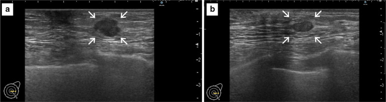
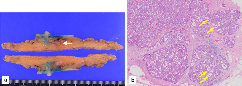
Case presentation: A 70-year-old man presented at our hospital with a primary complaint of pain and a lump in his left breast. Ultrasonography revealed a hypoechoic lesion measuring 12 mm × 10 mm × 8 mm. Fine-needle aspiration cytology revealed numerous necrotic and degenerated cells and few sheet-like clusters of atypical ductal epithelial cells. The atypical cells had mildly enlarged nuclei with nucleoli, were focally overlapped and formed tubular patterns. The cytological diagnosis indicated a suspicion of malignancy. Core needle biopsy (CNB) revealed necrotic and degenerated cells with microcalcification. The pathological diagnosis was indeterminate because there was no area of viable atypical cells. An excisional biopsy of the left breast lesion was scheduled one month later. However, it was difficult to detect the tumor during physical examination and ultrasonography performed 1 month after the patient's first visit. The operation was canceled, and the patient received follow-up observation. After 8 months of follow-up, ultrasonography and computed tomography (CT) revealed reappearance of a 0.6-cm-diameter breast tumor in the same place. CNB was performed again and revealed invasive ductal carcinoma. A total mastectomy with sentinel lymph node biopsy was performed 13 months after the first tumor disappeared. Histopathological examination revealed invasive cribriform carcinoma without sentinel lymph node metastasis. The patient did not have any complications, and adjuvant therapy with tamoxifen was started. The patient was alive without recurrence 7 months after surgery.
Conclusions: Temporary SR followed by tumor reappearance can occur in breast cancer cases, and it is important to follow patients even if their breast tumor has seemingly disappeared. When breast tumors disappear without treatment, clinicians must be aware of the possibility of SR of cancer and should follow the patient for early detection of tumor reappearance.
Keywords: Breast cancer; Cribriform; Reappearance; Spontaneous regression.
Conflict of interest statement
The authors declare that they have no competing interests.
Figures


References
LinkOut - more resources
Full Text Sources
Research Materials
