. 2020 Dec 7;18(12):e3001023.
doi: 10.1371/journal.pbio.3001023. eCollection 2020 Dec.

Prior expectations evoke stimulus-specific activity in the deep layers of the primary visual cortex

Affiliations

Prior expectations evoke stimulus-specific activity in the deep layers of the primary visual cortex

Fraser Aitken et al. PLoS Biol. .

Abstract

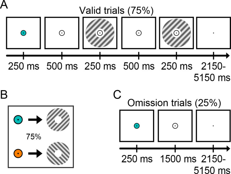
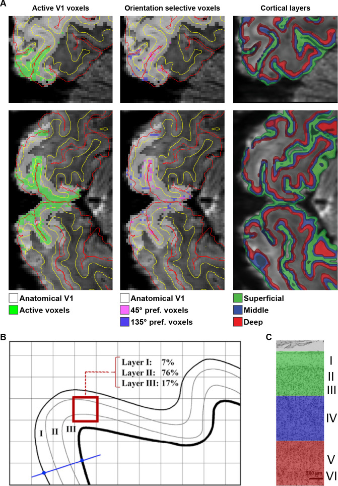
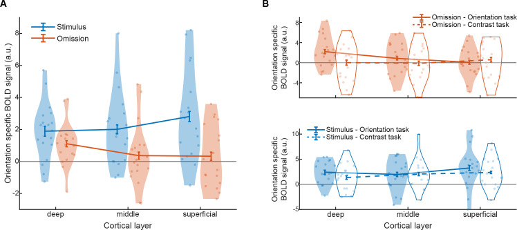
The way we perceive the world is strongly influenced by our expectations. In line with this, much recent research has revealed that prior expectations strongly modulate sensory processing. However, the neural circuitry through which the brain integrates external sensory inputs with internal expectation signals remains unknown. In order to understand the computational architecture of the cortex, we need to investigate the way these signals flow through the cortical layers. This is crucial because the different cortical layers have distinct intra- and interregional connectivity patterns, and therefore determining which layers are involved in a cortical computation can inform us on the sources and targets of these signals. Here, we used ultra-high field (7T) functional magnetic resonance imaging (fMRI) to reveal that prior expectations evoke stimulus-specific activity selectively in the deep layers of the primary visual cortex (V1). These findings are in line with predictive processing theories proposing that neurons in the deep cortical layers represent perceptual hypotheses and thereby shed light on the computational architecture of cortex.


Conflict of interest statement

The authors have declared that no competing interests exist.

Figures

Fig 1
Fig 1. Experimental paradigm.
(A) Each trial started with a coloured dot that predicted the orientation of the subsequent grating stimulus (45° or 135°). On 75% of trials, a set of gratings was then presented, the first of which had the expected orientation and the second differed slightly in orientation and contrast. In separate experimental runs, participants discriminated either the orientation or the contrast difference between the gratings. (B) The colour of the fixation circle (cyan or orange) predicted the orientation of the subsequent grating stimulus (45° or 135°) with 75% validity. (C) In the remaining 25% of trials, the gratings were omitted. On these trials, there was an expectation of a particular visual stimulus but no actual visual input. Participants had no task in these trials, except for holding central fixation.
Fig 2
Fig 2. Analysis approach.
(A) Illustration of ROI selection on sagittal slice of the mean functional scan of 1 participant. Overlaid red and yellow lines indicate pial and WM boundaries, respectively, as determined by registering anatomical boundaries to the mean functional image using RBR. Within V1 (white), active voxels were selected based on significant activation in the functional grating localiser (green). From these active voxels, we selected the 500 most strongly 45°-preferring (pink) and 135°-preferring (blue) voxels, respectively. With all voxels in these 2 ROIs, we determined how their volume was distributed over the superficial, middle, and deep cortical layers. (B) Schematic example of a voxel (red square) and the distribution of its volume over the 3 GM layers. This layer volume distribution was determined for each voxel and used as the basis of a regression approach in order to obtain layer-specific BOLD time courses (see Materials and methods). (C) Deep, middle, and superficial cortical layers indicated in coloured ribbons. Cytoarchitectural image of V1 adapted from [88]. BOLD, blood oxygen level–dependent; GM, grey matter; RBR, recursive boundary registration; ROI, region of interest; V1, primary visual cortex; WM, white matter.
Fig 3
Fig 3. Layer-specific BOLD response in V1 for presented and expected stimuli.
(A) Orientation-specific BOLD response to presented (blue) and expected-but-omitted (orange) gratings in the different layers of V1, averaged over tasks. (B) Orientation-specific BOLD response to expected-but-omitted (orange, top panel) and presented (blue, bottom panel) gratings, separately for the orientation (solid lines, filled shapes) and contrast (dashed lines, open shapes) tasks. Dots represent individual participants, and curved shapes indicate density. Error bars indicate within-subject SEM. Data are available at osf.io/k54p3. BOLD, blood oxygen level–dependent; SEM, standard error of the mean; V1, primary visual cortex.

References

    1. Kok P, Jehee JFM, De Lange FP. Less Is More: Expectation Sharpens Representations in the Primary Visual Cortex. Neuron. 2012;75:265–270. 10.1016/j.neuron.2012.04.034 - DOI - PubMed
    1. Yon D, Gilbert SJ, de Lange FP, Press C. Action sharpens sensory representations of expected outcomes. Nat Commun. 2018;9:4288 10.1038/s41467-018-06752-7 - DOI - PMC - PubMed
    1. Alink A, Schwiedrzik CM, Kohler A, Singer W, Muckli L. Stimulus Predictability Reduces Responses in Primary Visual Cortex. J Neurosci. 2010;30:2960–2966. 10.1523/JNEUROSCI.3730-10.2010 - DOI - PMC - PubMed
    1. Summerfield C, Trittschuh EH, Monti JM, Mesulam M-M, Egner T. Neural repetition suppression reflects fulfilled perceptual expectations. Nat Neurosci. 2008;11:1004–1006. 10.1038/nn.2163 - DOI - PMC - PubMed
    1. Todorovic A, van Ede F, Maris E, de Lange FP. Prior Expectation Mediates Neural Adaptation to Repeated Sounds in the Auditory Cortex: An MEG Study. J Neurosci. 2011;31:9118–9123. 10.1523/JNEUROSCI.1425-11.2011 - DOI - PMC - PubMed

Publication types