. 2020 Dec 3;12(12):3620.
doi: 10.3390/cancers12123620.

CNS Invasion in Meningioma-How the Intraoperative Assessment Can Improve the Prognostic Evaluation of Tumor Recurrence

Affiliations

CNS Invasion in Meningioma-How the Intraoperative Assessment Can Improve the Prognostic Evaluation of Tumor Recurrence

Felix Behling et al. Cancers (Basel). .

Abstract

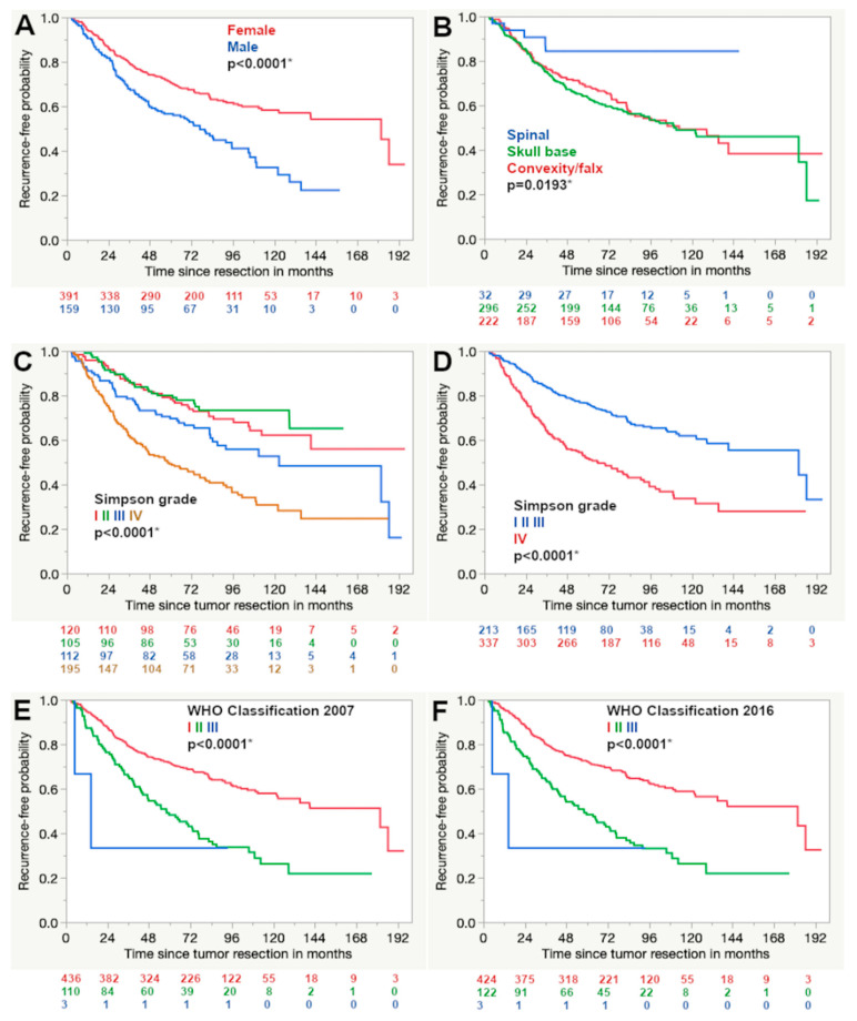
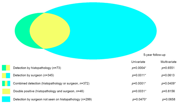
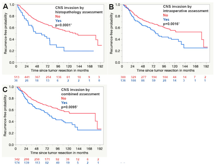
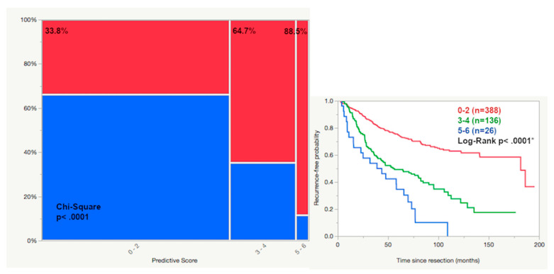
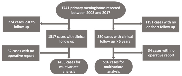
The detection of the infiltrative growth of meningiomas into CNS tissue has been integrated into the WHO classification as a stand-alone marker for atypical meningioma. However, its prognostic impact has been questioned. Infiltrative growth can also be detected intraoperatively. The prognostic impact of the intraoperative detection of the central nervous system tissue invasion of meningiomas was analyzed and compared to the histopathological assessment. The clinical data of 1517 cases with follow-up data regarding radiographic recurrence was collected. Histopathology and operative reports were reviewed and invasive growth was seen during resection in 23.7% (n = 345) while histopathology detected it in 4.8% (n = 73). The histopathological and intraoperative assessments were compatible in 63%. The prognostic impact of histopathological and intraoperative assessment was significant in the univariate but not in the multivariate analysis. Both methods of assessment combined reached statistical significance in the multivariate analysis (p = 0.0409). A score including all independent prognostic factors divided the cohort into three prognostic subgroups with a risk of recurrence of 33.8, 64.7 and 88.5%, respectively. The intraoperative detection of the infiltrative growth of primary meningiomas into the central nervous system tissue can complement the histopathological assessment of CNS invasion. The combined assessment is an independent prognostic factor regarding tumor recurrence and allows a risk-adapted tumor stratification.

Keywords: CNS invasion; brain invasion; intraoperative assessment; invasive growth; meningioma; progression-free survival; recurrence risk.


Conflict of interest statement

The authors declare no conflict of interest.

Figures

Figure 1
Figure 1
Kaplan–Meier curves show the results of the univariate survival analysis. A shorter progression-free survival was detected for male patients (A), non-spinal tumor location (B), higher Simpson (C,D) and WHO grade according to the classifications of 2007 and 2016 (E,F, respectively) (Log rank test, asterisk (*): statistically significant result).
Figure 2
Figure 2
Graphical depiction of the different subgroups of primary meningiomas with CNS invasion that were analyzed. The color represents the mode of assessment: histopathology (green and yellow), intraoperative (yellow and blue), combined histopathology and intraoperative (green, yellow and blue), double positive by histopathology and intraoperative (yellow), only positive by intraoperative assessment (blue). All subgroups were significant factors for tumor recurrence in the univariate analysis, but only the combined assessment (histopathology and intraoperative) turned out to be an independent significant prognostic factor in the multivariate analysis (asterisk (*): statistically significant result).
Figure 3
Figure 3
Kaplan–Meier curves demonstrate that the detection of CNS invasion by histopathology (A) and intraoperative assessment (B) were associated with a shorter progression-free survival. The combination of both detection methods was also a significant factor (C) (Log rank test, asterisk (*): statistically significant result).
Figure 4
Figure 4
The predictive score consists of the sum of the following factors: male gender (1), CNS invasion detected by histopathology or intraoperative assessment (1), WHO grade II or III (2) and Simpson grade >3 (2). Scoring groups of 0–2, 3–4 and 5–6 were generated and showed a significant difference in tumor recurrence of 33.8, 64.7 and 88.5%, respectively (recurrence red, non-recurrence blue, 5-year follow-up or longer, asterisk (*): statistically significant result).
Figure 5
Figure 5
Flow chart of the study cohort.

References

    1. Ostrom Q.T., Cioffi G., Gittleman H., Patil N., Waite K., Kruchko C., Barnholtz-Sloan J.S. CBTRUS Statistical Report: Primary Brain and Other Central Nervous System Tumors Diagnosed in the United States in 2012–2016. Neuro. Oncol. 2019;21 doi: 10.1093/neuonc/noz150. - DOI - PMC - PubMed
    1. Goldbrunner R., Minniti G., Preusser M., Jenkinson M.D., Sallabanda K., Houdart E., von Deimling A., Stavrinou P., Lefranc F., Lund-Johansen M., et al. EANO guidelines for the diagnosis and treatment of meningiomas. Lancet Oncol. 2016;17 doi: 10.1016/S1470-2045(16)30321-7. - DOI - PubMed
    1. McCarthy B.J., Davis F.G., Freel S., Surawicz T.S., Damek D.M., Grutsch J., Menck H.R., Laws E.R., Jr. Factors associated with survival in patients with meningioma. J. Neurosurg. 1998;88:831–839. doi: 10.3171/jns.1998.88.5.0831. - DOI - PubMed
    1. Louis D.N., Perry A., Reifenberger G., von Deimling A., Figarella-Branger D., Cavenee W.K., Ohgaki H., Wiestler O.D., Kleihues P., Ellison D.W. The 2016 World Health Organization Classification of Tumors of the Central Nervous System: A summary. Acta Neuropathol. 2016;131:803–820. doi: 10.1007/s00401-016-1545-1. - DOI - PubMed
    1. Brokinkel B., Hess K., Mawrin C. Brain invasion in meningiomas-clinical considerations and impact of neuropathological evaluation: A systematic review. Neuro. Oncol. 2017;19:1298–1307. doi: 10.1093/neuonc/nox071. - DOI - PMC - PubMed

LinkOut - more resources