. 2020 Dec 3;9(12):2595.
doi: 10.3390/cells9122595.

Focused Ultrasound-Induced Cavitation Sensitizes Cancer Cells to Radiation Therapy and Hyperthermia

Affiliations

Focused Ultrasound-Induced Cavitation Sensitizes Cancer Cells to Radiation Therapy and Hyperthermia

Shaonan Hu et al. Cells. .

Abstract

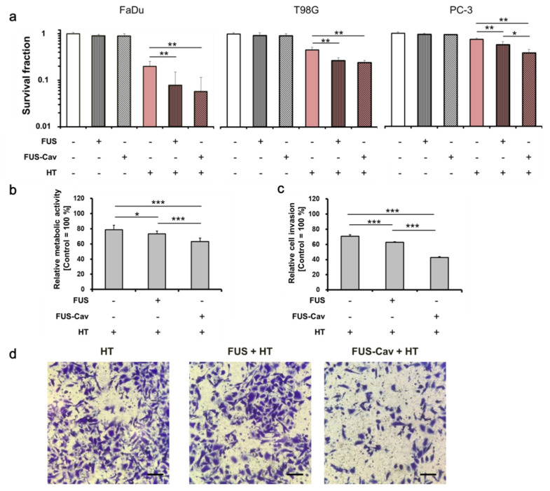
Focused ultrasound (FUS) has become an important non-invasive therapy for solid tumor ablation via thermal effects. The cavitation effect induced by FUS is thereby avoided but applied for lithotripsy, support drug delivery and the induction of blood vessel destruction for cancer therapy. In this study, head and neck cancer (FaDu), glioblastoma (T98G), and prostate cancer (PC-3) cells were exposed to FUS by using an in vitro FUS system followed by single-dose X-ray radiation therapy (RT) or water bath hyperthermia (HT). Sensitization effects of short FUS shots with cavitation (FUS-Cav) or without cavitation (FUS) to RT or HT (45 °C, 30 min) were evaluated. FUS-Cav significantly increases the sensitivity of cancer cells to RT and HT by reducing long-term clonogenic survival, short-term cell metabolic activity, cell invasion, and induction of sonoporation. Our results demonstrated that short FUS treatment with cavitation has good potential to sensitize cancer cells to RT and HT non-invasively.

Keywords: cavitation; focused ultrasound; hyperthermia; radiation therapy; sensitization.


Conflict of interest statement

The authors declare that they have no competing interests.

Figures

Figure 1
Figure 1
Experimental setup for cavitation detection and focused ultrasound (FUS) treatment. The in vitro FUS system includes a signal generator (Agilent 33120A), a radio frequency (RF) power amplifier (Electronics and Innovation A075), a customized FUS transducer and a water tank with adjustable heater and pump. (a) For the cavitation measurement, a 96-well plate filled with water was immersed in the water tank, the fiber-optic hydrophone (FOH) sensor was placed close to the bottom of the plate at the focal point acquiring the cavitation signal with oscilloscope. (b) FUS treatment was performed with monolayer cancer cells in a 96-well plate, the plate was sealed with water-proofed film to keep cells in a sterilized environment, a thermal camera (Optris PI450) was utilized for real-time temperature monitoring. The FUS treatment was controlled with a feedback loop to the thermal camera using LabView program.
Figure 2
Figure 2
Experimental timeline describing the procedure of the combination treatments and biological assays. The combination treatment of FUS/FUS-Cav/ hyperthermia (HT) and radiation therapy (RT) is shown in the blue square, and the treatment process of the combination of FUS/FUS-Cav and HT is shown in the red square.
Figure 3
Figure 3
Representative acoustic emission signals at 1.467 MHz and influence of acoustic intensity on the type and dose of cavitation. (a) The time-domain plots of the acoustic signals for one recording period of 2.9 s. (b) Corresponding frequency-domain plots; green arrow: fundamental frequency (f0) 1.467 MHz; blue arrow: sub- and ultra-harmonics (f = m × f0 /2, m = 1, 3, 5…) represent stable cavitation; red arrow: broadband noise represent inertial cavitation. (c) Root-mean-square (RMS) voltage of the broadband noise as a function of exposure time. (d) Stable and (e) inertial cavitation dose with a sonication duration of 40 s measured with a fiber-optic hydrophone, n = 9. (f) Fluorescence intensity measured by the terephthalic acid (TA) method indicates an inertial cavitation dose with a sonication duration of 40 s, n = 9.
Figure 4
Figure 4
FUS (129 W/cm2, 40 s) and FUS-Cav (1136 W/cm2, 40 s) revealed a radioadditive effect on reproductive long-term survival, metabolic activity and cell invasion. (a) Temperature curves of FUS and FUS-Cav treatment with a mean temperature of 36.99 ± 1.67 °C and 36.50 ± 1.53 °C. The diagram illustrated the sonication active duration (red arrow) and idle (black arrow) duration in each cycle, and the total treatment duration of FUS and FUS-Cav is 73.7 s and 126.7 s, respectively. (b) The clonogenic survival of FaDu, T98G and PC-3 cells was evaluated after FUS and FUS-Cav treatment in combination with radiation at a single dose of 5 or 10 Gy. Water bath HT (45 °C, 30 min) was performed as a reference. (c) Relative cell metabolic activity of PC-3 cells measured with WST-1 assay and (d) semi-quantitative result of the Transwell® assay indicating cell invasive potential of PC-3 cells 48 h post-treatment. Data were normalized to untreated control, which were set as 100% and relative values presented as mean ± standard error of the mean (SEM), n = 6, * p ≤ 0.05; ** p ≤ 0.01; *** p ≤ 0.001. (e) Representative microscopy images of Transwell® assay in PC-3 cells 48 h post-treatment. Scale bar = 100 µm.
Figure 5
Figure 5
FUS (129 W/cm2, 40 s) or FUS-Cav (1136 W/cm2, 40 s) demonstrated the additive effect to HT (45 °C, 30 min). (a) Clonogenic survival of FaDu, T98G, and PC-3 cells was evaluated after HT, FUS, and FUS-Cav treatment. (b) Relative cell metabolic activity of PC-3 cells measured with WST-1 assay 48 h post-treatment. (c) Semi-quantitative result of the Transwell® assay indicating cell invasive potential of PC-3 cells. (d) Representative microscopy images of Transwell® assay 48 h post-treatment. Scale bar = 100 µm. Data were normalized to the untreated control, which were set as 100% and relative values presented as mean ± SEM, n = 6, * p ≤ 0.05; ** p ≤ 0.01; *** p ≤ 0.001.
Figure 6
Figure 6
FUS-Cav induces sonoporation. FaDu and PC-3 cells were treated by FUS-Cav treatment at 1136 W/cm2 and with propidium iodide (PI) as a marker during and 30 min after treatment. (a) Representative fluorescence microscopy images showed an increase in red PI fluorescence during FUS-Cav. PI-stained cell nucleus (red) and CellMask-stained cell membrane (green), scale bar = 30 µm. (b) Semi-quantitative result of PI-positive percentage representing the occurrence of sonoporation. Data normalized to total cell number as 100%, n = 6, * p ≤ 0.05; ** p ≤ 0.01; *** p ≤ 0.001.

References

    1. Siedek F., Yeo S.Y., Heijman E., Grinstein O., Bratke G., Heneweer C., Puesken M., Persigehl T., Maintz D., Grüll H. Magnetic Resonance-Guided High-Intensity Focused Ultrasound (MR-HIFU): Technical Background and Overview of Current Clinical Applications (Part 1) RöFo—Fortschr. Geb. Röntgenstrahlen Bildgeb. Verfahr. 2019;191:522–530. doi: 10.1055/a-0817-5645. - DOI - PubMed
    1. Bour P., Ozenne V., Marquet F., De Senneville B.D., Dumont E., Quesson B. Real-time 3D ultrasound based motion tracking for the treatment of mobile organs with MR-guided high-intensity focused ultrasound. Int. J. Hyperth. 2018;34:1225–1235. doi: 10.1080/02656736.2018.1433879. - DOI - PubMed
    1. Elming P.B., Sørensen B.S., Oei A.L., Franken N.A., Crezee J., Overgaard J., Horsman M.R. Hyperthermia: The Optimal Treatment to Overcome Radiation Resistant Hypoxia. Cancers. 2019;11:60. doi: 10.3390/cancers11010060. - DOI - PMC - PubMed
    1. Roti J.L.R. Introduction: Radiosensitization by hyperthermia. Int. J. Hyperth. 2004;20:109–114. doi: 10.1080/0265673032000173898. - DOI - PubMed
    1. Peeken J.C., Vaupel P., Combs S.E. Integrating Hyperthermia into Modern Radiation Oncology: What Evidence Is Necessary? Front. Oncol. 2017;7:7. doi: 10.3389/fonc.2017.00132. - DOI - PMC - PubMed

Publication types