Review
. 2020 Dec 3;21(23):9238.
doi: 10.3390/ijms21239238.

Mitochondria and Calcium Homeostasis: Cisd2 as a Big Player in Cardiac Ageing

Affiliations
Review

Mitochondria and Calcium Homeostasis: Cisd2 as a Big Player in Cardiac Ageing

Chi-Hsiao Yeh et al. Int J Mol Sci. .

Abstract

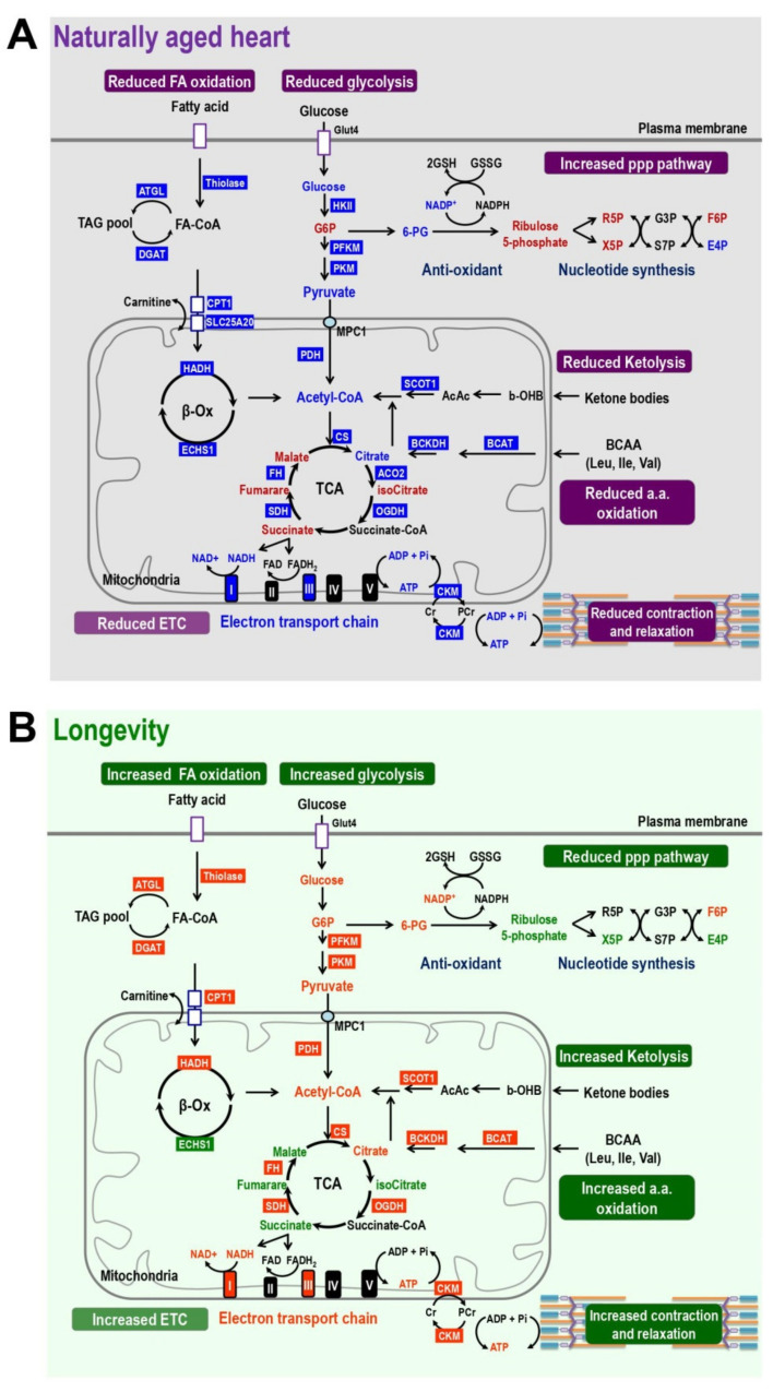
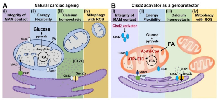
The ageing of human populations has become a problem throughout the world. In this context, increasing the healthy lifespan of individuals has become an important target for medical research and governments. Cardiac disease remains the leading cause of morbidity and mortality in ageing populations and results in significant increases in healthcare costs. Although clinical and basic research have revealed many novel insights into the pathways that drive heart failure, the molecular mechanisms underlying cardiac ageing and age-related cardiac dysfunction are still not fully understood. In this review we summarize the most updated publications and discuss the central components that drive cardiac ageing. The following characters of mitochondria-related dysfunction have been identified during cardiac ageing: (a) disruption of the integrity of mitochondria-associated membrane (MAM) contact sites; (b) dysregulation of energy metabolism and dynamic flexibility; (c) dyshomeostasis of Ca2+ control; (d) disturbance to mitochondria-lysosomal crosstalk. Furthermore, Cisd2, a pro-longevity gene, is known to be mainly located in the endoplasmic reticulum (ER), mitochondria, and MAM. The expression level of Cisd2 decreases during cardiac ageing. Remarkably, a high level of Cisd2 delays cardiac ageing and ameliorates age-related cardiac dysfunction; this occurs by maintaining correct regulation of energy metabolism and allowing dynamic control of metabolic flexibility. Together, our previous studies and new evidence provided here highlight Cisd2 as a novel target for developing therapies to promote healthy ageing.

Keywords: Cisd2; calcium homeostasis; cardiac ageing; energy flexibility; mitochondria; mitochondria-associated membranes (MAMs).


Conflict of interest statement

The authors declare no conflict of interest.

Figures

Figure 1
Figure 1
Cisd2 delays cardiac ageing as revealed by reduced lipofuscin accumulation and preserved myocardial ultrastructure. (A) Lipofuscin deposits can be clearly identified in cardiac tissues by autofluorescence detected with 330–380 nm excitation light and 420 nm barrier filter of a confocal fluorescence microscope. The red color indicates lipofuscin and green color indicates myocardia. (B) There is a significant increase of lipofuscin accumulation during cardiac ageing of WT mice. Intriguingly, a high level of Cisd2 reduces lipofuscin accumulation in Cisd2TG mice, while Cisd2 deficiency significantly elevates lipofuscin accumulation in Cisd2KO mice. The areas of lipofuscin accumulation and the myocardia are measured using Fiji/ImageJ 1.52e (National Institutes of Health, Bethesda, MD, USA). Ten fields per subject are observed at 20× objective magnification, and the lipofuscin accumulation (lipofuscin deposit area/myocardial area) was calculated. * p < 0.05, ** p < 0.01, *** p < 0.001. (C,D) Transmission electron microscopy (TEM) analysis for the left ventricle of heart. In 24-mo WT mice (C), age-associated mitochondria degeneration, lipofuscin accumulation, and necrotic debris of degenerative myofibril and organelles are detectable and easily identified. (D) In 24-mo Cisd2TG mice, relatively normal ultrastructures, namely intact mitochondria, ER, and cardiac myofibrils, are found. (C′) (D′) Schematic presentation for ultrastructure of left ventricle shown in (C) and (D), respectively. MD: mitochondrial degeneration, Lf: lipofuscin, ND: necrotic debris of degenerative myofibril and organelles, G: Golgi apparatus, M: mitochondria, Cm: cardiac myofibril, ER: endoplasmic reticulum, N: nucleus.
Figure 2
Figure 2
Cisd2 preserves the integrity of subsarcolemmal mitochondria and maintains the ultrastructure of mitochondria-associated ER membrane (MAM). (A) Cisd2 preserves the integrity of subsarcolemmal mitochondria and cardiac myofibrils of left ventricle. In 3-mo WT mice, there is an abundance of normal subsarcolemmal mitochondria and normal cardiac myofibrils (A-1 and A-1′). In 3-mo Cisd2KO mice (prematurely aged), notably, a low density of subsarcolemmal mitochondria, and degeneration of mitochondria and myofibrils are found (A-2 and A-2′). In 24-mo WT mice (naturally aged), similar ultrastructural damages are found as those observed in the 3-mo Cisd2KO mice (A-3 and A-3′). Remarkably, in 24-mo Cisd2TG mice, a high level of Cisd2 preserves the density and integrity of subsarcolemmal mitochondria as well as maintains the ultrastructure of cardiac myofibrils (A-4 and A-4′). (A-1′) to (A-4′) are schematic presentations for ultrastructure of left ventricle shown in (A-1) to (A-4). SL: sarcolemma, IS: intercellular space, M: mitochondria, Cm: cardiac myofibril, ICD: intercalated disc, MD: mitochondrial degeneration, Cmd: cardiac myofibril degeneration. (B) Cisd2 is essential to maintaining the integrity of MAM. In 3-mo WT mice, at MAM, mitochondria closely attach to ER (B-1 and B-1′). In 3-mo Cisd2KO mice, notably, a longer distance between mitochondria and ER is found; in addition, mitochondria degeneration is also found (B-2 and B-2′). In 24-mo WT mice, a longer distance between mitochondria and ER is found; this is similar to that observed in the 3-mo Cisd2KO mice (B-3 and B-3′). Intriguingly, in 24-mo Cisd2TG mice, a high level of Cisd2 maintains a relatively normal MAM, where the mitochondria closely attach to the ER (B-4 and B-4′). (B-1′) to (B-4′) are schematic presentations for the ultrastructure of left ventricle shown in (B-1) to (B-4). The inset shows a higher magnification to provide a better illustration for MAM. The inset shows a higher magnification of the selected area (yellow squares) of the middle panel to provide a better illustration for MAM.
Figure 3
Figure 3
Cisd2 maintains a younger pattern of energy metabolism which is dysregulated in naturally aged heart. (A) Energy metabolism is disrupted during natural aging of wild-type (WT) mice. The results are obtained by comparing 24-mo WT vs. 3-mo WT mice. In the aged heart, the TCA cycle and ATP production are impaired due to decrease of fatty acid oxidation, ketolysis, glucose utilization, and amino acid oxidation. In addition, the creatinine shuttling of high-energy phosphate is also diminished in the aged heart. Blue and red colors mark the enzymes and metabolites that are downregulated or upregulated in the pathways, respectively. (B) Cisd2 maintains a younger pattern of energy metabolism in the heart of Cisd2TG mice, which is a long-lived mouse model. The results are obtained by comparing 24-mo Cisd2TG vs. 24-mo WT mice. Remarkably, in the Cisd2TG heart, there is an increase in the fatty acid oxidation, ketolysis, glucose utilization, and amino acid oxidation. Consequently, this appears to result in a normal cycling of the TCA and an increased activity of the electron transport chain to produce ATP. In addition, the creatinine shuttling of high-energy phosphate is also restored in the longevity heart. All together, these beneficial effects brought about by Cisd2 provide a sufficient energy supply to fuel the heart of the Cisd2TG mice. The levels of the enzymes and metabolites which have an opposite profile to that observed in the naturally aged heart are marked by green (down) and orange (up). In this study, alterations of metabolic pathways are summarized from omics analyses of metabolomics and transcriptomics of RNA sequencing using cardiac tissues of left ventricles.
Figure 4
Figure 4
Cisd2 preserves the structure and function of MAM during cardiac ageing. (A) Four characters of mitochondria-related dysfunction are identified during cardiac ageing: (i) Disruption of integrity of MAM contact sites. Physical proximity and functional interplay between mitochondria and SR are disturbed during cardiac ageing; this is associated with a widened gap distance between ER and mitochondria leading to an abnormal Ca2+ signaling and lipid transport. (ii) Dysregulation of energy flexibility. More than 95% of cardiac ATP is produced from the oxidative phosphorylation of mitochondria via electron transfer chain (ETC), which is fueled with energy primarily from β-oxidation of fatty acids (FA) and to a lesser extent from glycolysis of glucose. Defective fatty acid oxidation of mitochondria jeopardizes the energy flexibility of cardiomyocytes. (iii) Ca2+ dyshomeostasis. Elevated cytosolic Ca2+ levels resulted from the defective Ca2+ recruitment of ER and decreased Ca2+ influx capacity of mitochondria as well as impaired cellular Ca2+ extrusions are detectable in aged hearts. (iv) Disturbance of mitophagy accompanied with increased ROS. Decrease in the capacity of lysosomal degradation pathway and mitochondrial biogenesis, as well as decrease in mitophagy are observed in aged hearts. Furthermore, these defects usually associated with increased oxidative stress. (B) Cisd2 activator as a geroprotector to increase Cisd2 and to preserve cardiac function during ageing. Cisd2 is mainly located in the ER, mitochondria, and MAM. The expression level of Cisd2 decreases during cardiac ageing. A high level of Cisd2 achieved by treatment of Cisd2 activators probably can attenuate these abnormalities during cardiac ageing.

References

    1. Virani S.S., Alonso A., Benjamin E.J., Bittencourt M.S., Callaway C.W., Carson A.P., Chamberlain A.M., Chang A.R., Cheng S., Delling F.N., et al. Heart Disease and Stroke Statistics-2020 Update: A Report from the American Heart Association. Circulation. 2020;141:e139–e596. doi: 10.1161/CIR.0000000000000757. - DOI - PubMed
    1. Li H., Hastings M.H., Rhee J., Trager L.E., Roh J.D., Rosenzweig A. Targeting age-related pathways in heart failure. Circ. Res. 2020;126:533–551. doi: 10.1161/CIRCRESAHA.119.315889. - DOI - PMC - PubMed
    1. Li J., Zhang D., Brundel B., Wiersma M. Imbalance of ER and mitochondria interactions: Prelude to cardiac ageing and disease? Cells. 2019;8:1617. doi: 10.3390/cells8121617. - DOI - PMC - PubMed
    1. Lakatta E.G. So! What’s aging? Is cardiovascular aging a disease? J. Mol. Cell Cardiol. 2015;83:1–13. doi: 10.1016/j.yjmcc.2015.04.005. - DOI - PMC - PubMed
    1. Lee K.-S., Huh S., Lee S., Wu Z., Kim A.-K., Kang H.-Y., Lu B. Altered ER-mitochondria contact impacts mitochondria calcium homeostasis and contributes to neurodegeneration in vivo in disease models. Proc. Natl. Acad. Sci. USA. 2018;115:E8844–E8853. doi: 10.1073/pnas.1721136115. - DOI - PMC - PubMed

LinkOut - more resources