

A collagen membrane influences bone turnover marker in vivo after bone augmentation with xenogenic bone
- PMID: 33287844
- PMCID: PMC7722310
- DOI: 10.1186/s13005-020-00249-9
A collagen membrane influences bone turnover marker in vivo after bone augmentation with xenogenic bone
Abstract
Background: The aim was to compare early biochemical and histological osseous healing of chronic mandibular defects regenerated with bovine bone substitute with and without collagen membrane in vivo.
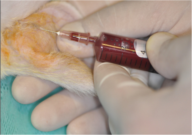
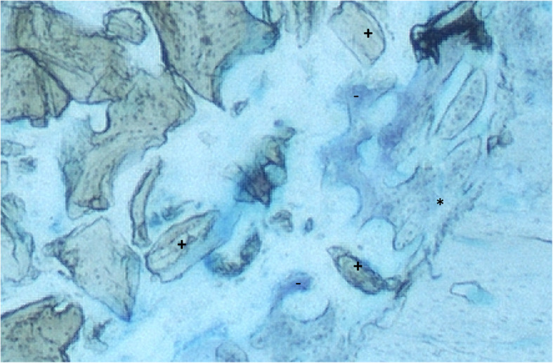
Methods: Eight weeks after formation of a lateral full-thickness perforating bone defect in the mandible of 40 rabbits, bovine bone substitute with ("+";n = 20) and without ("-";n = 20) collagen membrane was applied. Blood and bone was collected 24, 72 h, 7, 14 and 21 days after surgery. Total acid phosphatase, bone acid phosphatase, total alkaline phosphatase and bone alkaline phosphatase activities were compared between groups. Formation of new bone was quantified histologically for all time points.
Results: Twenty-four hours after surgery, bone alkaline phosphatase was significantly elevated in "+" group when compared to "-" (p=0.012). After 72 hours, all bone turnover markers except for total acid phosphatase (p=0.078) where significantly elevated in "+" (all p < 0.05). Fourteen days after surgery, the significant highest values for all bone turnover markers were detected in "-" (all p < 0.05). A significant difference in favor of group "-" could also be detected after 3 weeks in terms of both acid phosphatases (p < 0.05). In histology, no significant differences could be detected.
Conclusion: Bone regeneration with bovine bone substitute material and collagen membrane shows a significantly earlier bone remodeling activity but does not seem to influence formation of new bone in histological samples.
Keywords: Animal study; Bone regeneration; Bone remodeling; Collagen; Membrane; Serological bone turnover markers.
Conflict of interest statement
The authors declare that they have no competing interests.
Figures


References
-
- Kämmerer PW, Palarie V, Schiegnitz E, Nacu V, Draenert FG, Al-Nawas B. Influence of a collagen membrane and recombinant platelet-derived growth factor on vertical bone augmentation in implant-fixed deproteinized bovine bone--animal pilot study. Clin Oral Implants Res. 2013;24(11):1222–1230. - PubMed
MeSH terms
Substances
LinkOut - more resources
Full Text Sources
