. 2020 Dec 22;117(51):32757-32763.
doi: 10.1073/pnas.2015550117. Epub 2020 Dec 7.

ARP2/3-independent WAVE/SCAR pathway and class XI myosin control sperm nuclear migration in flowering plants

Affiliations

ARP2/3-independent WAVE/SCAR pathway and class XI myosin control sperm nuclear migration in flowering plants

Mohammad Foteh Ali et al. Proc Natl Acad Sci U S A. .

Abstract

After eukaryotic fertilization, gamete nuclei migrate to fuse parental genomes in order to initiate development of the next generation. In most animals, microtubules control female and male pronuclear migration in the zygote. Flowering plants, on the other hand, have evolved actin filament (F-actin)-based sperm nuclear migration systems for karyogamy. Flowering plants have also evolved a unique double-fertilization process: two female gametophytic cells, the egg and central cells, are each fertilized by a sperm cell. The molecular and cellular mechanisms of how flowering plants utilize and control F-actin for double-fertilization events are largely unknown. Using confocal microscopy live-cell imaging with a combination of pharmacological and genetic approaches, we identified factors involved in F-actin dynamics and sperm nuclear migration in Arabidopsis thaliana (Arabidopsis) and Nicotiana tabacum (tobacco). We demonstrate that the F-actin regulator, SCAR2, but not the ARP2/3 protein complex, controls the coordinated active F-actin movement. These results imply that an ARP2/3-independent WAVE/SCAR-signaling pathway regulates F-actin dynamics in female gametophytic cells for fertilization. We also identify that the class XI myosin XI-G controls active F-actin movement in the Arabidopsis central cell. XI-G is not a simple transporter, moving cargos along F-actin, but can generate forces that control the dynamic movement of F-actin for fertilization. Our results provide insights into the mechanisms that control gamete nuclear migration and reveal regulatory pathways for dynamic F-actin movement in flowering plants.

Keywords: F-actin; WAVE/SCAR; fertilization; myosin; nuclear migration.


Conflict of interest statement

The authors declare no competing interest.

Figures

Fig. 1.
Fig. 1.
F-actin dynamics in the central cell is WAVE/SCAR dependent, but ARP2/3 complex independent. (A, Top) Z-projected confocal image of the central cell F-actin (cyan, proFWA::Lifeact:Venus), central cell nucleus (yellow, proFWA::H2B:mRuby2), and autofluorescence (magenta) marking the central cell border. (A, Bottom) Schematic diagram of the mature Arabidopsis ovule. Arrows indicate the direction of central cell F-actin movement from the plasma membrane periphery to the nucleus. (BD, F, and G) Time-lapse (1-min interval, marked by five different colors) stacks of Z-projected central cell F-actin images of the mock treatment (B), wiskostatin (10 µM for 1h incubation) treatment (C), scar2-1 mutant (D), CK-666 (200 µM for 1 h incubation) treatment (F), and the arp2-1 mutant (G). Dashed circles indicate the position of the central cell nucleus. F-actin marked by different colors denotes F-actin movement, whereas white color resulting from overlapping of all colors represents less or no movement. (E) The transcriptional activity of the Arabidopsis ARP2 promoter is visualized by proARP2::H2B:Clover (green). Arrow points to the central cell nucleus and autofluorescence marks the central cell border. (H) Mean velocity of F-actin dynamics in the central cell (**P < 0.001; ns, not significant; Tukey-Kramer HSD test). The box spans first and third quartiles, and the line inside the box shows the median. Bars on the top and bottom represent the maximum and minimum values. (Scale bar, 20 µm.)
Fig. 2.
Fig. 2.
Sperm nuclear migration is delayed in scar2-1 and xi-g central cells. (AD) Representative images of sperm chromatin dynamics: (A) Sperm cells just released from the pollen tube into the ovule. (B) Sperm nuclei starting to move toward the central cell and egg cell nuclei. (C) Sperm chromatin decondensed in both the central cell and egg-cell nuclei. (D) Delayed karyogamy and sperm chromatin decondensation observed in the scar2-1 central cell. Note that sperm chromatin became fully decondensed in the egg cell nucleus while sperm chromatin remained condensed in the central cell. This delayed phenotype was not observed in WT. Sperm chromatin was visualized by the sperm-specific histone marker proHTR10::HTR10:mRFP1. Arrows and arrowheads point to the condensed and decondensed sperm chromatin, respectively. Dashed circles indicate the position of the central cell nucleus. Autofluorescence of the central cell border was also visualized. (E) Status of sperm chromatin 9 h after pollination. Stages AD are shown in AD, respectively. (Scale bar, 20 µm.)
Fig. 3.
Fig. 3.
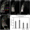
The class XI myosin XI-G is involved in F-actin meshwork movement in the Arabidopsis central cell. (AD) Time-lapse (1-min interval, marked by five different colors) stacks of Z-projected central cell F-actin images of the mock treatment (A), the xi-g mutant (B), 20 mM BDM treatment (C), and 50 mM BDM treatment (D). Dashed circles indicate the position of the central cell nucleus. F-actin marked by different colors denotes F-actin movement, whereas white color resulting from overlapping of all colors, represents less or no movement. (E) The transcriptional activity of the Arabidopsis XI-G promoter is visualized by proXI-G::H2B:Clover (green). Autofluorescence marks the central cell wall; the arrow and arrowheads point to the central cell nucleus and synergid nuclei, respectively. (F) Mean velocity of F-actin dynamics in the central cell. Levels not connected by the same letter (a–c) are significantly different (P < 0.01, Tukey-Kramer HSD test). The box spans first and third quartiles, and the line inside the box shows the median. Bars on the top and bottom represent the maximum and minimum values. (G) The orientation of F-actin in the central cell was evaluated by measuring the angles of F-actin cables (shown in red) made with a line radiating from the center of the central cell nucleus (shown as dashed lines). Black dots represent individual angle data and violin shapes show the kernel probability densities (**P < 0.001; Tukey-Kramer HSD test). (Scale bar, 20 µm.)
Fig. 4.
Fig. 4.
The 20 mM BDM affects mitochondrial movement in the Arabidopsis central cell. (AD) Time-lapse (1-min interval, marked by five different colors) stacks of Z-projected central cell mitochondrial movement images of the mock treatment (A), 20 mM BDM treatment (B), 50 mM BDM treatment (C), and in the xi-g mutant (D). Mitochondria marked by different colors denote movement, whereas white color resulting from overlapping of all colors represents less or no movement. Dashed circles indicate the position of the central cell nucleus. (E) Average velocity of mitochondrial movement in the central cell. Error bars represent SEM. Levels not connected by the same letter (A and B) are significantly different (P < 0.01, Tukey-Kramer HSD test). (Scale bar, 20 µm.)
Fig. 5.
Fig. 5.
Model of F-actin dynamics in the female gamete for sperm nuclear migration. (A and B) Schematic image of F-actin meshwork movement (A) and the pathway controlling F-actin movement in the Arabidopsis central cell (B). ROP8 localizes to the plasma membrane and interacts with SCAR2. The ROP8-SCAR2–signaling pathway positively regulates the meshwork F-actin movement in a gametophyte-specific ARP2/3-independent manner. Myosins including XI-G also regulate the meshwork F-actin movement through myosin functions distinguishable from the movement of organelles.

References

    1. Kawashima T., Berger F., Green love talks: Cell-cell communication during double fertilization in flowering plants. AoB Plants 2011, plr015 (2011). - PMC - PubMed
    1. Shin J. M., Yuan L., Ohme-Takagi M., Kawashima T., Cellular dynamics of double fertilization and early embryogenesis in flowering plants. J. Exp. Zoolog. B Mol. Dev. Evol., 10.1002/jez.b.22981 (2020). - DOI - PubMed
    1. Reinsch S., Gönczy P., Mechanisms of nuclear positioning. J. Cell Sci. 111, 2283–2295 (1998). - PubMed
    1. Fatema U., Ali M. F., Hu Z., Clark A. J., Kawashima T., Gamete nuclear migration in animals and plants. Front. Plant Sci. 10, 517 (2019). - PMC - PubMed
    1. Kawashima T., et al. , Dynamic F-actin movement is essential for fertilization in Arabidopsis thaliana. eLife 3, e04501 (2014). - PMC - PubMed

Publication types

MeSH terms

LinkOut - more resources