. 2020 Dec 22;117(51):32370-32379.
doi: 10.1073/pnas.2016158117. Epub 2020 Dec 7.

Mesyl phosphoramidate backbone modified antisense oligonucleotides targeting miR-21 with enhanced in vivo therapeutic potency

Affiliations

Mesyl phosphoramidate backbone modified antisense oligonucleotides targeting miR-21 with enhanced in vivo therapeutic potency

Olga A Patutina et al. Proc Natl Acad Sci U S A. .

Abstract

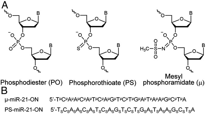
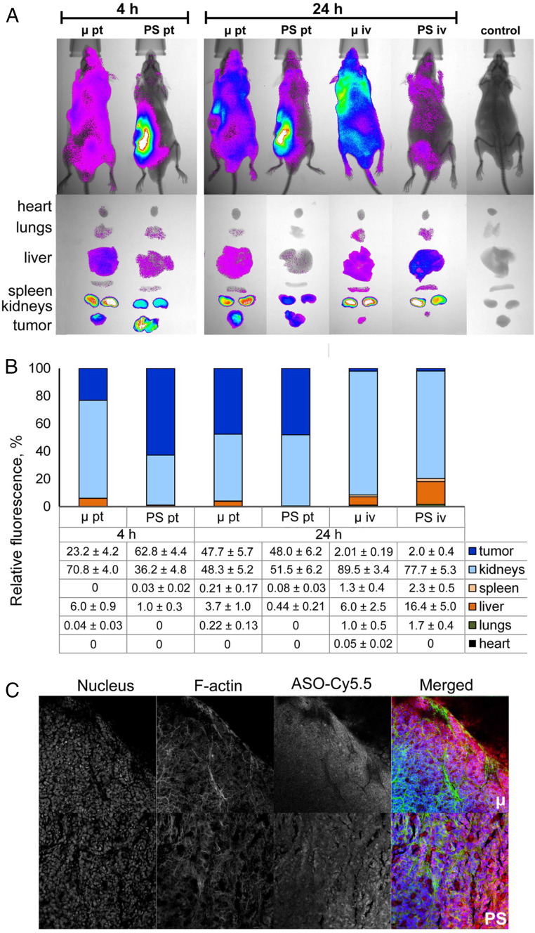
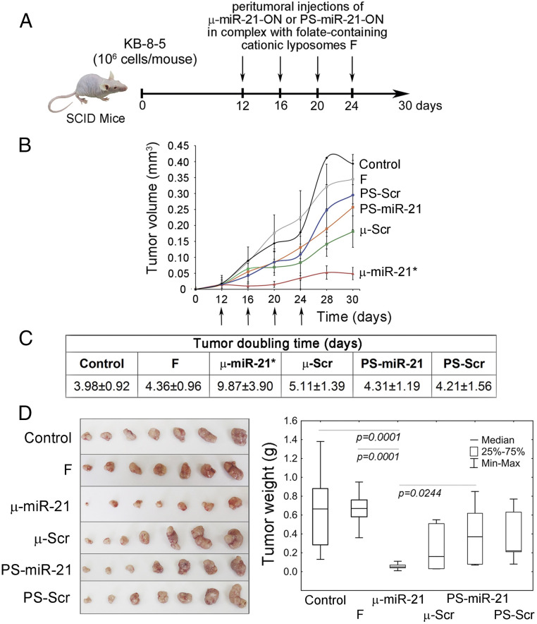
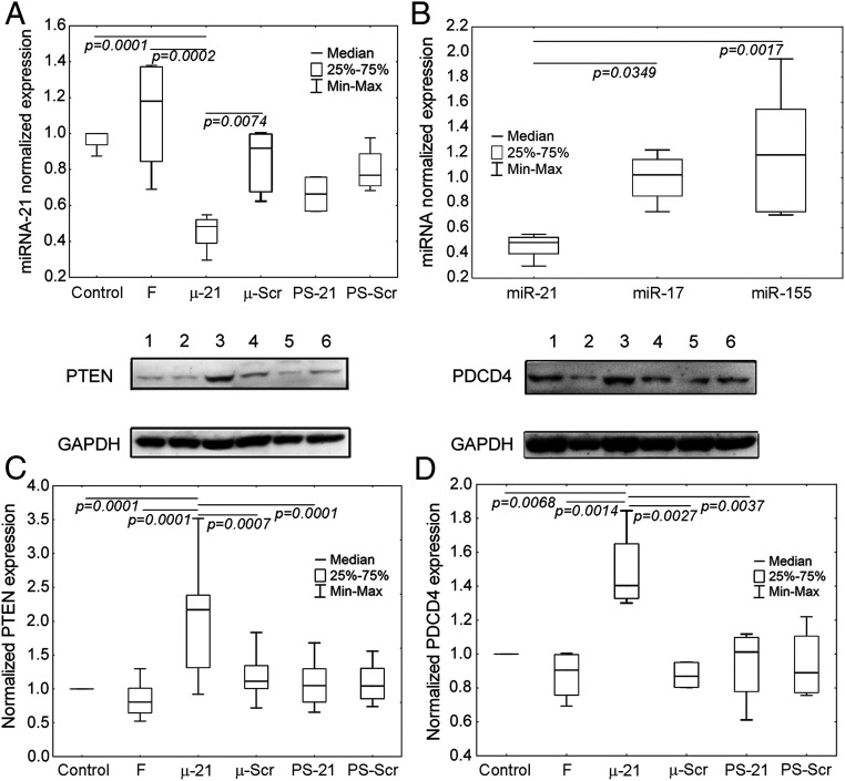
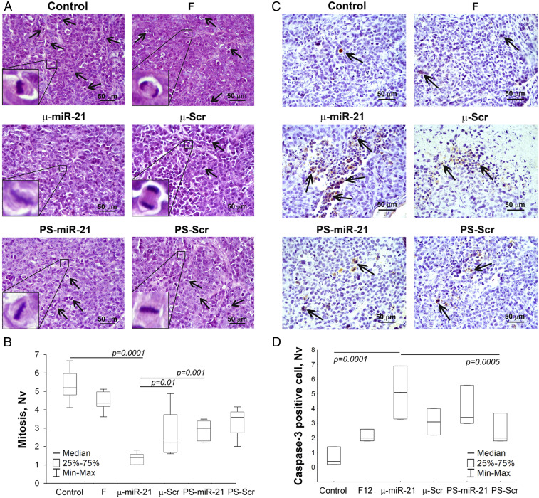
The design of modified oligonucleotides that combine in one molecule several therapeutically beneficial properties still poses a major challenge. Recently a new type of modified mesyl phosphoramidate (or µ-) oligonucleotide was described that demonstrates high affinity to RNA, exceptional nuclease resistance, efficient recruitment of RNase H, and potent inhibition of key carcinogenesis processes in vitro. Herein, using a xenograft mouse tumor model, it was demonstrated that microRNA miR-21-targeted µ-oligonucleotides administered in complex with folate-containing liposomes dramatically inhibit primary tumor growth via long-term down-regulation of miR-21 in tumors and increase in biosynthesis of miR-21-regulated tumor suppressor proteins. This antitumoral effect is superior to the effect of the corresponding phosphorothioate. Peritumoral administration of µ-oligonucleotide results in its rapid distribution and efficient accumulation in the tumor. Blood biochemistry and morphometric studies of internal organs revealed no pronounced toxicity of µ-oligonucleotides. This new oligonucleotide class provides a powerful tool for antisense technology.

Keywords: DNA modification; antisense oligonucleotide; mesyl oligonucleotide; oncogenic microRNA; phosphorothioate.


Conflict of interest statement

The authors declare no competing interest.

Figures

Fig. 1.
Fig. 1.
Structures and sequences of antisense oligonucleotides (ASOs) targeted to miR-21. (A) Structures of DNA phosphate group modifications: phosphodiester (PO), phosphorothioate (PS), and mesyl phosphoramidate (μ). (B) Sequences of oligonucleotides μ-miR-21-ON and PS-miR-21-ON used in the study. Indices μ and S stand for mesyl phosphoramidate group and phosphorothioate group, respectively.
Fig. 2.
Fig. 2.
Biodistribution of Cy5.5-labeled µ and PS ASOs in tumor-bearing SCID mice. (A) Lifetime fluorescence imaging of tumor-bearing SCID mice (n = 3 mice per group), dorsal orientation view, and dissected organs 4 and 24 h after peritumoral (pt) and i.v. (iv) injections of 40 µg per mouse of modified oligonucleotides. Control, noninjected mice. (B) Percent biodistribution of modified oligonucleotides after 4 and 24 h after pt and i.v. injections of µ and PS ASOs (mean ± SE). (C) Confocal microscopic images of cryosections of KB-8-5 tumors from mice 24 h after pt injections of Cy5.5-labeled μ and PS ASOs at a dose of 40 μg per mouse. Three-channel pictures are shown. Merged images represent staining of nucleus with DAPI (blue), staining of actin filaments with phalloidin-TRITC (green), and visualization of oligonucleotides by Cy5.5-labeling (red). Magnification ×400.
Fig. 3.
Fig. 3.
Antitumor effect of µ-miR-21-ON. (A) Experimental design including engraftment of SCID mice with human epidermoid carcinoma KB-8-5 and peritumoral injections of µ-miR-21-ON, PS-miR-21-ON, or scrambled µ or PS ASOs in complex with folate-containing liposomes F at a dose of 10 µg per mouse. In total, four injections were made at days 12, 16, 20, and 24 after tumor cell implantation. (B) Kinetics of KB-8-5 tumor growth after treatment with µ-miR-21-ON and PS-miR-21-ON (n = 7 mice per group). The days of injections are marked by arrows. An * indicates statistically significant difference of μ-miR-21-ON group from all of the other groups, with P < 0.05. (C) Doubling time of KB-8-5 tumors after treatment with oligonucleotides. Statistically reliable difference from all groups is marked by asterisk. (D) Tumor weight at day 30. Data were statistically analyzed using one-way ANOVA with post hoc Tukey test; P value indicates a statistically reliable difference. F, liposomes F.
Fig. 4.
Fig. 4.
The level of miRNAs and proteins PTEN and PDCD4 in human epidermoid carcinoma KB-8-5 tissue after administration of μ-miR-21-ON and PS-miR-21-ON. (A) The level of miR-21 in tumor tissue after completion of treatment with μ-miR-21-ON and PS-miR-21-ON. (B) The level of miR-21, miR-155, and miR-17 after treatment with μ-miR-21-ON. Expression of miRNAs was measured by qPCR and normalized to the expression of snRNA U6. (C and D) Expression of tumor suppressor proteins PTEN and PDCD4, respectively, after treatment with μ-miR-21-ON and PS-miR-21-ON measured by Western blot. Protein levels were normalized to the level of GAPDH. Numbers indicate the following groups: 1, control; 2, folate-containing liposomes F; 3, μ-miR-21-ON; 4, μ-Scr-ON; 5, PS-miR-21-ON; 6, Ps-Scr-ON. Data were statistically analyzed using one-way ANOVA with post hoc Tukey test; P value indicates a statistically reliable difference.
Fig. 5.
Fig. 5.
Mitosis and apoptosis in KB-8-5 tumors after therapy with µ-miR-21-ON and PS-miR-21-ON. (A) Typical images of tumor sections after hematoxylin and eosin staining. Magnification ×400. Bar corresponds to 50 µm. Mitoses are indicated by arrows. Typical examples of individual mitotic events are shown with magnification ×1,000 in the bottom left corner. (B) Morphometric analysis of tumor tissue with mitosis counting. (C) Typical images of tumor sections after immunohistochemical staining with caspase-3 monoclonal antibodies. Examples of caspase-3–positive cells are indicated by arrows. Magnification ×400. (D) Morphometric analysis of tumor tissue with apoptotic cells counting. Nv ×400. Nv, the numerical density indicating the number of particles in the unit tissue volume. Data were statistically analyzed using one-way ANOVA with post hoc Tukey test; P value indicates a statistically reliable difference.

References

    1. Crooke S. T., Witztum J. L., Bennett C. F., Baker B. F., RNA-targeted therapeutics. Cell Metab. 27, 714–739 (2018). - PubMed
    1. Shen X., Corey D. R., Chemistry, mechanism and clinical status of antisense oligonucleotides and duplex RNAs. Nucleic Acids Res. 46, 1584–1600 (2018). - PMC - PubMed
    1. Eckstein F., Phosphorothioates, essential components of therapeutic oligonucleotides. Nucleic Acid Ther. 24, 374–387 (2014). - PubMed
    1. Shen W., et al. , Chemical modification of PS-ASO therapeutics reduces cellular protein-binding and improves the therapeutic index. Nat. Biotechnol. 37, 640–650 (2019). - PubMed
    1. Migawa M. T., et al. , Site-specific replacement of phosphorothioate with alkyl phosphonate linkages enhances the therapeutic profile of gapmer ASOs by modulating interactions with cellular proteins. Nucleic Acids Res. 47, 5465–5479 (2019). - PMC - PubMed

Publication types

MeSH terms

LinkOut - more resources