. 2020 Dec 7;3(1):740.
doi: 10.1038/s42003-020-01479-y.

Epigenetic landscape of pancreatic neuroendocrine tumours reveals distinct cells of origin and means of tumour progression

Affiliations

Epigenetic landscape of pancreatic neuroendocrine tumours reveals distinct cells of origin and means of tumour progression

Annunziata Di Domenico et al. Commun Biol. .

Abstract

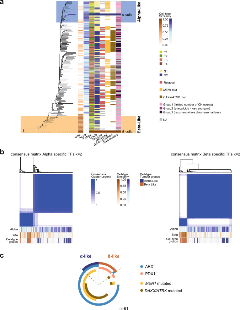
Recent data suggest that Pancreatic Neuroendocrine Tumours (PanNETs) originate from α- or β-cells of the islets of Langerhans. The majority of PanNETs are non-functional and do not express cell-type specific hormones. In the current study we examine whether tumour DNA methylation (DNAme) profiling combined with genomic data is able to identify cell of origin and to reveal pathways involved in PanNET progression. We analyse genome-wide DNAme data of 125 PanNETs and sorted α- and β-cells. To confirm cell identity, we investigate ARX and PDX1 expression. Based on epigenetic similarities, PanNETs cluster in α-like, β-like and intermediate tumours. The epigenetic similarity to α-cells progressively decreases in the intermediate tumours, which present unclear differentiation. Specific transcription factor methylation and expression vary in the respective α/β-tumour groups. Depending on DNAme similarity to α/β-cells, PanNETs have different mutational spectra, stage of the disease and prognosis, indicating potential means of PanNET progression.


Conflict of interest statement

The authors declare no competing interests.

Figures

Fig. 1
Fig. 1. PanNET methylomes resemble distinct endocrine cell lineages.
a Phyloepigenetic analysis of PanNET (n = 125) and normal α- and β-cell samples (n = 2 for each cell type). A rooted tree was created with an arbitrary chosen β-sample as the root and selecting the differentially methylated CpGs between sorted normal α- and β-cell samples (n = 2131, adj. p value < 0.001 and |Δβ | > 0.2). Blue and orange scales show the degree of similarity of each PanNET sample to α- and β-cells, respectively. The degree of similarity was calculated via DNA-methylation cell-type deconvolution comparing tumour methylomes to sorted normal α- and β-cell methylomes. Each line represents a patient. Clinical and molecular features for each sample are indicated. b Consensus clustering of the 125 PanNETs according to the α- and β-cell-specific TF-checkpoint methylation sites (49 CpGs for β-cells and 51 CpGs for α-cells). The k mean value was set to 2. Consensus cluster correlation is indicated according to the blue scale as depicted. Fractions of similarity to α- and β-cells for each sample are reported at the bottom of each matrix according to the blue and orange scales. Similarly, the cell-type-specific tumour groups are reported for each sample in blue (α-like), orange (β-like), and white (intermediate). c Schematic overview of the PanNET subtypes. Doughnut chart for all samples showing cell-specific PanNET subtypes, ARX and PDX1 expression, MEN1 and DAXX/ATRX mutations.
Fig. 2
Fig. 2. Expression of ARX, PDX1, insulin, and glucagon can be used as surrogate for defining tumour cell-type specificity.
a Immunostaining for the different subtypes of PanNETs. From the top to the bottom: tumour positive for ARX and glucagon; tumour positive for ARX; double positive tumour for PDX1 and ARX; double negative tumour for ARX and PDX1; tumour positive for PDX1 and insulin. For the TFs only nuclear staining was considered for scoring. b Venn diagram displaying the total number of samples positive for ARX and/or PDX1or negative for both the transcription factors. c Schematic overview of the PanNET subtypes in cohort 2. Doughnut chart for all samples showing glucagon, insulin, PDX1 and ARX positivity as well as loss of DAXX/ATRX expression.
Fig. 3
Fig. 3. Epigenetic differentiation status defines clinically different PanNETs and draws two possible ways of PanNET evolution.
a Kaplan–Meier disease free survival of 95 patients (cohort 1) stratified according to cell-type specific methylation groups (α-like in blue, β-like in orange and intermediate in grey). Intermediate tumours have higher risk of relapse compared to α- and β-like tumours (p value = 0.00016). In b and c disease free survival of a subset of patients of cohort 1 (n = 50) with available data for both DNA-methylation profile and PDX1, ARX, and DAXX/ATRX IHC. In b patient stratification according to cell-type-specific methylation groups and in c according to PDX1, ARX, and DAXX/ATRX IHC (in grey patients double negative for PDX1 and ARX or ARX positive and negative for DAXX/ATRX, in light-blue patients double negative for PDX1 and ARX or ARX positive and DAXX/ATRX positive, in light-orange patients double positive for PDX1 and ARX or PDX1 positive and DAXX/ATRX positive). d Consensus clustering of the 125 PanNETs according to the 6364 differentially methylated sites between α-like, β-like and intermediate tumours (adj. p value < 0.001 and |Δβ| > 0.2). Cluster stability was reached for k = 4 (see Supplementary Fig. 7a). Consensus cluster correlation is indicated according to the blue scale as depicted. Each column represents one PanNET sample. Tumour mutations and cell-type subgroups are indicated according to the reported colours. e Progression model hypothesis based on epigenetic and genetic evolution: α-like tumours originate from α-cells upon MEN1 inactivation, the progression to intermediate-ADM tumours is enhanced by secondary events, including loss of DAXX/ATRX and chromosomal instability with recurrent LOH and activation of ALT. These events are associated to a gradual loss of differentiation. Beta-like tumours originate from β-cells upon different genetic event and they are mainly insulinoma, usually indolent. However, based on our data it is not possible to exclude that intermediate-WT PanNETs originate directly from β-cells or from a progenitor cell. Similarly we cannot exclude that intermediate-ADM may originate from a precursor cell as well. Progression supported by genetic and epigenetic data is depicted by full lines, while hypothetical progression is depicted by dotted lines.

References

    1. Boninsegna L, et al. Malignant pancreatic neuroendocrine tumour: lymph node ratio and Ki67 are predictors of recurrence after curative resections. Eur. J. Cancer. 2012;48:1608–1615. doi: 10.1016/j.ejca.2011.10.030. - DOI - PubMed
    1. Falconi M, et al. ENETS consensus guidelines update for the management of patients with functional pancreatic neuroendocrine tumors and non-functional pancreatic neuroendocrine tumors. Neuroendocrinology. 2016;103:153–171. doi: 10.1159/000443171. - DOI - PMC - PubMed
    1. Di Domenico A, Wiedmer T, Marinoni I, Perren A. Genetic and epigenetic drivers of neuroendocrine tumours (NET) Endocr. Relat. Cancer. 2017;24:R315–R334. doi: 10.1530/ERC-17-0012. - DOI - PubMed
    1. Marinoni I, et al. Loss of DAXX and ATRX are associated with chromosome instability and reduced survival of patients with pancreatic neuroendocrine tumors. Gastroenterology. 2014;146:453–460.e5. doi: 10.1053/j.gastro.2013.10.020. - DOI - PubMed
    1. Sadanandam A, et al. A cross-species analysis in pancreatic neuroendocrine tumors reveals molecular subtypes with distinctive clinical, metastatic, developmental, and metabolic characteristics. Cancer Discov. 2015;5:1296–1313. doi: 10.1158/2159-8290.CD-15-0068. - DOI - PMC - PubMed

Publication types

MeSH terms