

Role of lung ultrasonography in the diagnosis of COVID-19 patients admitted to the emergency department
- PMID: 33288981
- PMCID: PMC7709804
- DOI: 10.1007/s10049-020-00807-3
Role of lung ultrasonography in the diagnosis of COVID-19 patients admitted to the emergency department
Abstract
Introduction: In this study, the use of lung ultrasonography (LUS) to diagnosis lung findings was evaluated in patients with suspected COVID-19 who were admitted to the emergency department (ED).
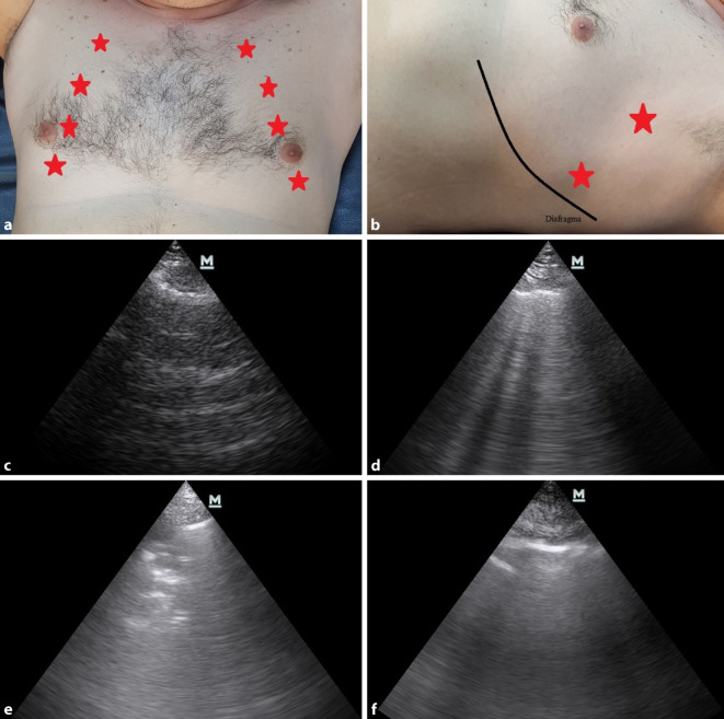
Methods: This observational clinical study was conducted in the ED of the Ankara City Hospital during the period April 1-30, 2020. Patients who were admitted to the ED were triaged as COVID-19 infected and who agreed to undergo LUS/LCT (lung computed tomography) were included in the study.
Results: Included in the study were 40 patients who had been prediagnosed with COVID-19. Pneumonia was detected with LCT in 32 (80%) patients, while the LUS examination identified pneumonia in 23 patients. The most common finding in LCT was ground-glass opacity (n = 29, 90.6%). Of the 23 patients with pneumonia findings in LUS, 15 (65.2%) had direct consolidation. Among the 32 patients who were found to have pneumonia as a result of LCT, 20 (62.5%) had signs of pneumonia on LUS examination, and 12 had no signs of pneumonia. In addition, 3 patients showed no signs of pneumonia with LCT, but they were misdiagnosed with pneumonia by LUS. The sensitivity of LUS in the diagnosis of pneumonia in the COVID-19 patients was 62.5%, while its specificity was 62.5%. In addition, its positive predictive value was 87.0%, and its negative predictive value was 29.4%.
Conclusion: LUS may also be used in the diagnosis of pneumonia in COVID-19 patients because it is a valuable and accessible bedside diagnostic tool.
Einführung: In dieser Studie wurde die Verwendung einer Lungenultraschalluntersuchung (LUS) zur Diagnose von Lungenbefunden bei Patienten, die mit Verdacht auf „coronavirus disease 19“ (COVID‑19) in die Notaufnahme (NA) aufgenommen worden sind, bewertet.
Methoden: Diese klinische Beobachtungsstudie wurde in der Notaufnahme des Stadtkrankenhaus Ankara in einem Zeitraum vom 1. bis 30. April 2020 durchgeführt. Patienten, die in der Notaufnahme als COVID-19-Infizierte erst eingeschätzt und Einwilligung für eine Lungenultraschalluntersuchung (LUS)/Lungencomputertomographie (LCT) gegeben haben, wurde in die Studie aufgenommen.
Ergebnisse: Insgesamt 40 mit COVID‑19 vordiagnostizierte Patienten wurden in die Studie eingeschlossen. Bei 32 Patienten (80 %) wurde anhand der CT eine Pneumonie festgestellt, während die LUS bei 23 Patienten eine Pneumonie feststellen konnte. Der häufigste Befund in der LCT war eine Milchglastrübung (n = 29, 90,6 %). Von den 23 Patienten mit Pneumoniebefund im LUS hatten 15 (65,2 %) eine direkte Konsolidierung. Von den 32 Patienten, bei denen die LCT einen Pneumoniebefund ergeben hatte, wiesen 20 (62,5 %) Anzeichen einer Pneumonie im LUS auf, bei 12 war dies nicht der Fall. Drei Patienten hatten keine Anzeichen einer Pneumonie in der LCT, wurden aber aufgrund der LUS mit einer Pneumonie fehldiagnostiziert. Die Sensitivität des LUS für die Diagnose einer Pneumonie bei COVID-19-Patienten betrug 62,5 %, die Spezifität 62,5 %. Der positive prädiktiver Wert betrug 87,0 %, der negative prädiktiver Wert 29,4 %.
Schlussfolgerungen: Der LUS kann auch in der Diagnostik einer Pneumonie bei COVID-19-Patienten eingesetzt werden, weil er ein wertvolles und zugängliches diagnostisches Instrument am Krankenbett ist.
Keywords: Consolidation; Diagnostic ultrasound; Emergency service, hospital; Pneumonia; SARS-CoV‑2.
© Springer Medizin Verlag GmbH, ein Teil von Springer Nature 2020.
Conflict of interest statement
Conflict of interestİ. Şan, B. Bekgöz, E. Usul, Ç. Yıldırım, E. Gemcioğlu, A.F. Kahraman and A.E. Ay declare that they have no competing interests.
Figures
References
-
- https://www.who.int/docs/default-source/coronaviruse/situation-reports/2.... Accessed 16 June 2020
-
- (2005) emergency-committee-regarding-the-outbreak-of-novel-coronavirus-(2019-ncov). https://www.who.int/news-room/detail/30-01-2020-statement-on-the-second-.... Accessed 16 June 2020
-
- https://www.who.int/docs/default-source/coronaviruse/situation-reports/2.... Accessed 17 June 2020
-
- World Health Organization (2020) Guidance W. Clinical management of severe acute respiratory infection when novel coronavirus (2019-nCoV) infection is suspected. WHO/2019-nCoV/clinical/2020.5
LinkOut - more resources
Full Text Sources
Miscellaneous