Fluorescence Guidance and Intraoperative Adjuvants to Maximize Extent of Resection
- PMID: 33289518
- PMCID: PMC8510852
- DOI: 10.1093/neuros/nyaa475
Fluorescence Guidance and Intraoperative Adjuvants to Maximize Extent of Resection
Abstract
Safely maximizing extent of resection has become the central goal in glioma surgery. Especially in eloquent cortex, the goal of maximal resection is balanced with neurological risk. As new technologies emerge in the field of neurosurgery, the standards for maximal safe resection have been elevated. Fluorescence-guided surgery, intraoperative magnetic resonance imaging, and microscopic imaging methods are among the most well-validated tools available to enhance the level of accuracy and safety in glioma surgery. Each technology uses a different characteristic of glioma tissue to identify and differentiate tumor tissue from normal brain and is most effective in the context of anatomic, connectomic, and neurophysiologic context. While each tool is able to enhance resection, multiple modalities are often used in conjunction to achieve maximal safe resection. This paper reviews the mechanism and utility of the major adjuncts available for use in glioma surgery, especially in tumors within eloquent areas, and puts forth the foundation for a unified approach to how leverage currently available technology to ensure maximal safe resection.
Keywords: 5-aminolevulinic acid (5-ALA); Fluorescence-guided surgery (FGS); Raman microscopy; intraoperative MRI (iMRI).
Copyright © 2020 by the Congress of Neurological Surgeons.
Figures


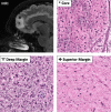
 ) revealed mildly gliotic cortex with normal cellularity without evidence of tumor infiltration. No additional resection was required or carried out at the superior margin.
Comment in
-
Letter: Fluorescence Guidance and Intraoperative Adjuvants to Maximize Extent of Resection.Neurosurgery. 2022 May 1;90(5):e137-e138. doi: 10.1227/neu.0000000000001916. Neurosurgery. 2022. PMID: 35238808 No abstract available.
-
In Reply: Fluorescence Guidance and Intraoperative Adjuvants to Maximize Extent of Resection.Neurosurgery. 2022 May 1;90(5):e139. doi: 10.1227/neu.0000000000001917. Neurosurgery. 2022. PMID: 35238813 Free PMC article. No abstract available.
References
-
- Lacroix M, Abi-Said D, Fourney DRet al. A multivariate analysis of 416 patients with glioblastoma multiforme: prognosis, extent of resection, and survival. J Neurosurg. 2001;95(2):190-198. - PubMed
-
- McGirt MJ, Chaichana KL, Gathinji Met al. Independent association of extent of resection with survival in patients with malignant brain astrocytoma: clinical article. J Neurosurg. 2009;110(1):156-162. - PubMed
-
- Bloch O, Han SJ, Cha Set al. Impact of extent of resection for recurrent glioblastoma on overall survival. J Neurosurg. 2012;117(6):1032-1038. - PubMed
-
- Fukui A, Muragaki Y, Saito Tet al. Volumetric analysis using low-field intraoperative magnetic resonance imaging for 168 newly diagnosed supratentorial glioblastomas: effects of extent of resection and residual tumor volume on survival and recurrence. World Neurosurg. 2017;98:73-80. - PubMed
-
- Coburger J, Merkel A, Scherer Met al. Low-grade glioma surgery in intraoperative magnetic resonance imaging: results of a multicenter retrospective assessment of the German Study Group for intraoperative magnetic resonance imaging. Neurosurgery. 2016;78(6):775-785. - PubMed
Publication types
MeSH terms
Substances
Grants and funding
LinkOut - more resources
Full Text Sources
Medical
Research Materials

