. 2021 Feb;298(2):365-373.
doi: 10.1148/radiol.2020200822. Epub 2020 Dec 8.

Improved Task-based Functional MRI Language Mapping in Patients with Brain Tumors through Marchenko-Pastur Principal Component Analysis Denoising

Affiliations

Improved Task-based Functional MRI Language Mapping in Patients with Brain Tumors through Marchenko-Pastur Principal Component Analysis Denoising

Benjamin Ades-Aron et al. Radiology. 2021 Feb.

Abstract

Background Functional MRI improves preoperative planning in patients with brain tumors, but task-correlated signal intensity changes are only 2%-3% above baseline. This makes accurate functional mapping challenging. Marchenko-Pastur principal component analysis (MP-PCA) provides a novel strategy to separate functional MRI signal from noise without requiring user input or prior data representation. Purpose To determine whether MP-PCA denoising improves activation magnitude for task-based functional MRI language mapping in patients with brain tumors. Materials and Methods In this Health Insurance Portability and Accountability Act-compliant study, MP-PCA performance was first evaluated by using simulated functional MRI data with a known ground truth. Right-handed, left-language-dominant patients with brain tumors who successfully performed verb generation, sentence completion, and finger tapping functional MRI tasks were retrospectively identified between January 2017 and August 2018. On the group level, for each task, histograms of z scores for original and MP-PCA denoised data were extracted from relevant regions and contralateral homologs were seeded by a neuroradiologist blinded to functional MRI findings. Z scores were compared with paired two-sided t tests, and distributions were compared with effect size measurements and the Kolmogorov-Smirnov test. The number of voxels with a z score greater than 3 was used to measure task sensitivity relative to task duration. Results Twenty-three patients (mean age ± standard deviation, 43 years ± 18; 13 women) were evaluated. MP-PCA denoising led to a higher median z score of task-based functional MRI voxel activation in left hemisphere cortical regions for verb generation (from 3.8 ± 1.0 to 4.5 ± 1.4; P < .001), sentence completion (from 3.7 ± 1.0 to 4.3 ± 1.4; P < .001), and finger tapping (from 6.9 ± 2.4 to 7.9 ± 2.9; P < .001). Median z scores did not improve in contralateral homolog regions for verb generation (from -2.7 ± 0.54 to -2.5 ± 0.40; P = .90), sentence completion (from -2.3 ± 0.21 to -2.4 ± 0.37; P = .39), or finger tapping (from -2.3 ± 1.20 to -2.7 ± 1.40; P = .07). Individual functional MRI task durations could be truncated by at least 40% after MP-PCA without degradation of clinically relevant correlations between functional cortex and functional MRI tasks. Conclusion Denoising with Marchenko-Pastur principal component analysis led to higher task correlations in relevant cortical regions during functional MRI language mapping in patients with brain tumors. © RSNA, 2020 Online supplemental material is available for this article.


Figures

None
Graphical abstract
(a) Plots of functional MRI signal intensity as a function of time (top [without denoising], bottom [with denoising]). We show mean normalized functional MRI signal intensity in the contralateral precentral gyrus hand knob during unilateral finger movements compared with signal intensity after denoising with Marchenko-Pastur principal component analysis (MP-PCA) for one patient. MP-PCA led to decreased random temporal fluctuations in the blood oxygenation level–dependent (BOLD) signal and increased correlation coefficient task model. (b) Plot of temporal correlation function as a function of lag time, f(Δt)/σ2 , of the signal residuals (denoised – original; see Materials and Methods) normalized by the estimated voxelwise noise level shows that residuals have no memory; its mean is zero for all lag times Δt except when Δt = 0, implying that no informative temporal correlations were removed during denoising.
Figure 1a:
(a) Plots of functional MRI signal intensity as a function of time (top [without denoising], bottom [with denoising]). We show mean normalized functional MRI signal intensity in the contralateral precentral gyrus hand knob during unilateral finger movements compared with signal intensity after denoising with Marchenko-Pastur principal component analysis (MP-PCA) for one patient. MP-PCA led to decreased random temporal fluctuations in the blood oxygenation level–dependent (BOLD) signal and increased correlation coefficient task model. (b) Plot of temporal correlation function as a function of lag time, ft)/σ2 , of the signal residuals (denoised – original; see Materials and Methods) normalized by the estimated voxelwise noise level shows that residuals have no memory; its mean is zero for all lag times Δt except when Δt = 0, implying that no informative temporal correlations were removed during denoising.
(a) Plots of functional MRI signal intensity as a function of time (top [without denoising], bottom [with denoising]). We show mean normalized functional MRI signal intensity in the contralateral precentral gyrus hand knob during unilateral finger movements compared with signal intensity after denoising with Marchenko-Pastur principal component analysis (MP-PCA) for one patient. MP-PCA led to decreased random temporal fluctuations in the blood oxygenation level–dependent (BOLD) signal and increased correlation coefficient task model. (b) Plot of temporal correlation function as a function of lag time, f(Δt)/σ2 , of the signal residuals (denoised – original; see Materials and Methods) normalized by the estimated voxelwise noise level shows that residuals have no memory; its mean is zero for all lag times Δt except when Δt = 0, implying that no informative temporal correlations were removed during denoising.
Figure 1b:
(a) Plots of functional MRI signal intensity as a function of time (top [without denoising], bottom [with denoising]). We show mean normalized functional MRI signal intensity in the contralateral precentral gyrus hand knob during unilateral finger movements compared with signal intensity after denoising with Marchenko-Pastur principal component analysis (MP-PCA) for one patient. MP-PCA led to decreased random temporal fluctuations in the blood oxygenation level–dependent (BOLD) signal and increased correlation coefficient task model. (b) Plot of temporal correlation function as a function of lag time, ft)/σ2 , of the signal residuals (denoised – original; see Materials and Methods) normalized by the estimated voxelwise noise level shows that residuals have no memory; its mean is zero for all lag times Δt except when Δt = 0, implying that no informative temporal correlations were removed during denoising.
Qualitative comparison of images obtained without denoising (Original) and with anisotropic smoothing, independent component analysis (ICA), denoising convolutional neural network (DnCNN), and Marchenko-Pastur principal component analysis (MP-PCA) denoising. MP-PCA shows a more spatially accurate activation pattern compared with smoothing and a greater volume of activation compared with other common image denoising routines. According to radiology conventions, the images show functional MRI changes after right-hand unilateral finger tapping overlaid on a 1-mm isotropic resolution magnetization-prepared rapid gradient-echo image in a patient with a left parietal glioblastoma and a threshold of z greater than 3.
Figure 2:
Qualitative comparison of images obtained without denoising (Original) and with anisotropic smoothing, independent component analysis (ICA), denoising convolutional neural network (DnCNN), and Marchenko-Pastur principal component analysis (MP-PCA) denoising. MP-PCA shows a more spatially accurate activation pattern compared with smoothing and a greater volume of activation compared with other common image denoising routines. According to radiology conventions, the images show functional MRI changes after right-hand unilateral finger tapping overlaid on a 1-mm isotropic resolution magnetization-prepared rapid gradient-echo image in a patient with a left parietal glioblastoma and a threshold of z greater than 3.
(a) Residual images, defined as the original image subtracted from modified versions of the image, for an individual performing the unilateral finger-tapping task. The four images differences are shown: smoothing – original, independent component analysis (ICA) – original, denoising convolutional neural network (DNCNN) – original, and Marchenko-Pastur principal component analysis (MPPCA) – original. Except for Marchenko-Pastur principal component analysis (MP-PCA), gyral brain structures are visible in the residual images. (b) Plot of the probability p(r) of removing noise at a given normalized variance level (r2). Normalized residual is defined as r = (denoised – original)/σ, where σ is the MP-PCA estimated noise level. Ideal denoising corresponds to removing Gaussian distributed residual, such that is a unit Gaussian (a straight black line in the log p(r) vs r2 plot). Histograms of residuals for all methods except MP-PCA exhibit strongly non-Gaussian residuals, indicating the removal of anatomic features. MP-PCA residuals are normally distributed down to an r2 almost equal to 12 (ie, 3.5 standard deviations in the tail of the Gaussian).
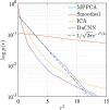
Figure 3a:
(a) Residual images, defined as the original image subtracted from modified versions of the image, for an individual performing the unilateral finger-tapping task. The four images differences are shown: smoothing – original, independent component analysis (ICA) – original, denoising convolutional neural network (DNCNN) – original, and Marchenko-Pastur principal component analysis (MPPCA) – original. Except for Marchenko-Pastur principal component analysis (MP-PCA), gyral brain structures are visible in the residual images. (b) Plot of the probability p(r) of removing noise at a given normalized variance level (r2). Normalized residual is defined as r = (denoised – original)/σ, where σ is the MP-PCA estimated noise level. Ideal denoising corresponds to removing Gaussian distributed residual, such that formula image is a unit Gaussian (a straight black line in the log p(r) vs r2 plot). Histograms of residuals for all methods except MP-PCA exhibit strongly non-Gaussian residuals, indicating the removal of anatomic features. MP-PCA residuals are normally distributed down to an r2 almost equal to 12 (ie, 3.5 standard deviations in the tail of the Gaussian).
(a) Residual images, defined as the original image subtracted from modified versions of the image, for an individual performing the unilateral finger-tapping task. The four images differences are shown: smoothing – original, independent component analysis (ICA) – original, denoising convolutional neural network (DNCNN) – original, and Marchenko-Pastur principal component analysis (MPPCA) – original. Except for Marchenko-Pastur principal component analysis (MP-PCA), gyral brain structures are visible in the residual images. (b) Plot of the probability p(r) of removing noise at a given normalized variance level (r2). Normalized residual is defined as r = (denoised – original)/σ, where σ is the MP-PCA estimated noise level. Ideal denoising corresponds to removing Gaussian distributed residual, such that is a unit Gaussian (a straight black line in the log p(r) vs r2 plot). Histograms of residuals for all methods except MP-PCA exhibit strongly non-Gaussian residuals, indicating the removal of anatomic features. MP-PCA residuals are normally distributed down to an r2 almost equal to 12 (ie, 3.5 standard deviations in the tail of the Gaussian).
Figure 3b:
(a) Residual images, defined as the original image subtracted from modified versions of the image, for an individual performing the unilateral finger-tapping task. The four images differences are shown: smoothing – original, independent component analysis (ICA) – original, denoising convolutional neural network (DNCNN) – original, and Marchenko-Pastur principal component analysis (MPPCA) – original. Except for Marchenko-Pastur principal component analysis (MP-PCA), gyral brain structures are visible in the residual images. (b) Plot of the probability p(r) of removing noise at a given normalized variance level (r2). Normalized residual is defined as r = (denoised – original)/σ, where σ is the MP-PCA estimated noise level. Ideal denoising corresponds to removing Gaussian distributed residual, such that formula image is a unit Gaussian (a straight black line in the log p(r) vs r2 plot). Histograms of residuals for all methods except MP-PCA exhibit strongly non-Gaussian residuals, indicating the removal of anatomic features. MP-PCA residuals are normally distributed down to an r2 almost equal to 12 (ie, 3.5 standard deviations in the tail of the Gaussian).
Z statistic maps from the sentence-completion functional MRI task for a representative patient (38-year-old man) with a glioblastoma in the posterior left middle temporal gyrus near receptive language regions. From left to right, the images shown are a magnetization-prepared rapid gradient-echo image with no overlay followed by the original activation map (z original), the denoised activation map (z denoised), and the areas of difference between denoised and original activation maps (difference).
Figure 4:
Z statistic maps from the sentence-completion functional MRI task for a representative patient (38-year-old man) with a glioblastoma in the posterior left middle temporal gyrus near receptive language regions. From left to right, the images shown are a magnetization-prepared rapid gradient-echo image with no overlay followed by the original activation map (z original), the denoised activation map (z denoised), and the areas of difference between denoised and original activation maps (difference).
Histograms of z score magnitude for the original data and Marchenko-Pastur principal component analysis (MP-PCA) denoised data in eight regions of interest (data are from all 23 patients): the left and right Broca area, the left and right hand knob, the left and right language or presupplementary motor area (SMA), and left and right Wernicke area. Vertical lines show distribution medians. Histogram outlines correspond to all voxels within the spherical regions of interest. Shaded histograms correspond to regions of interest based on the first principal component. Histogram outlines are normalized to have an area under the curve of 1 standard deviation, and shaded histograms are normalized to the number of voxels included in the respective region of interest. Shaded areas in the right hemisphere and in motor regions are small because there are fewer highly correlated voxels in these areas compared with the left hemisphere language–dominant regions. Denoising widens the tails of these distributions, and in areas where task-related activation is increased, there is an outward shift in distribution median. There are shifts in the opposite direction in the right hemisphere due to task lateralization in the brain in contralateral regions.
Figure 5:
Histograms of z score magnitude for the original data and Marchenko-Pastur principal component analysis (MP-PCA) denoised data in eight regions of interest (data are from all 23 patients): the left and right Broca area, the left and right hand knob, the left and right language or presupplementary motor area (SMA), and left and right Wernicke area. Vertical lines show distribution medians. Histogram outlines correspond to all voxels within the spherical regions of interest. Shaded histograms correspond to regions of interest based on the first principal component. Histogram outlines are normalized to have an area under the curve of 1 standard deviation, and shaded histograms are normalized to the number of voxels included in the respective region of interest. Shaded areas in the right hemisphere and in motor regions are small because there are fewer highly correlated voxels in these areas compared with the left hemisphere language–dominant regions. Denoising widens the tails of these distributions, and in areas where task-related activation is increased, there is an outward shift in distribution median. There are shifts in the opposite direction in the right hemisphere due to task lateralization in the brain in contralateral regions.
Graphs show task sensitivity (measured by using the percentage of voxels in the whole brain with |z| > 3) as a function of total imaging time, on average for all 23 patients (shaded areas are the population standard deviations). With Marchenko-Pastur principal component analysis (MP-PCA) denoising, patients performing the verb-generation and unilateral finger-tapping task will reach the same number of voxels with z greater than 3 by using only 60% of the original imaging time, whereas the sentence-completion task can be reduced by 50%.
Figure 6:
Graphs show task sensitivity (measured by using the percentage of voxels in the whole brain with |z| > 3) as a function of total imaging time, on average for all 23 patients (shaded areas are the population standard deviations). With Marchenko-Pastur principal component analysis (MP-PCA) denoising, patients performing the verb-generation and unilateral finger-tapping task will reach the same number of voxels with z greater than 3 by using only 60% of the original imaging time, whereas the sentence-completion task can be reduced by 50%.

Comment in

References

    1. Pillai JJ. The evolution of clinical functional imaging during the past 2 decades and its current impact on neurosurgical planning. AJNR Am J Neuroradiol 2010;31(2):219–225. - PMC - PubMed
    1. Petrella JR, Shah LM, Harris KM, et al. Preoperative functional MR imaging localization of language and motor areas: effect on therapeutic decision making in patients with potentially resectable brain tumors. Radiology 2006;240(3):793–802. - PubMed
    1. Hoult DI, Lauterbur PC. The sensitivity of the zeugmatographic experiment involving human samples. J Magn Reson (1969) 1979;34(2):425–433.
    1. Welvaert M, Rosseel Y. On the definition of signal-to-noise ratio and contrast-to-noise ratio for FMRI data. PLoS One 2013;8(11):e77089. - PMC - PubMed
    1. Zacà D, Nickerson JP, Deib G, Pillai JJ. Effectiveness of four different clinical fMRI paradigms for preoperative regional determination of language lateralization in patients with brain tumors. Neuroradiology 2012;54(9):1015–1025. - PubMed

Publication types

LinkOut - more resources