. 2021 Mar;298(3):E131-E140.
doi: 10.1148/radiol.2020203496. Epub 2020 Dec 8.

Utility of Screening Chest Radiographs in Patients with Asymptomatic or Minimally Symptomatic COVID-19 in Singapore

Affiliations

Utility of Screening Chest Radiographs in Patients with Asymptomatic or Minimally Symptomatic COVID-19 in Singapore

Benjamin J Kuo et al. Radiology. 2021 Mar.

Abstract

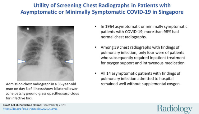
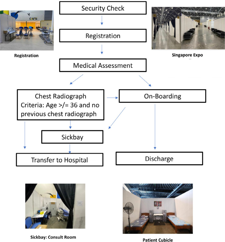
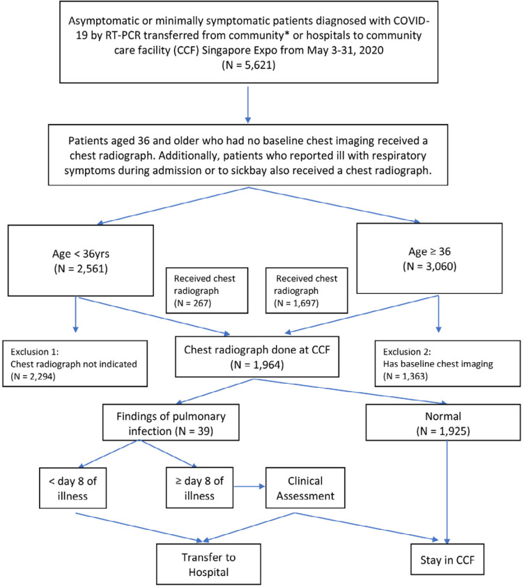
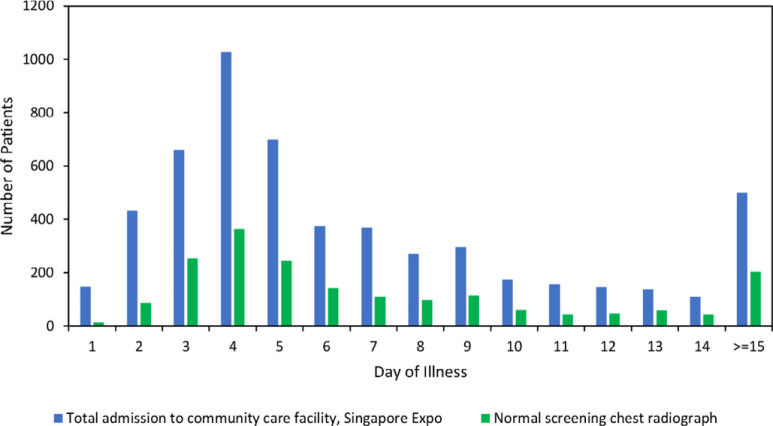
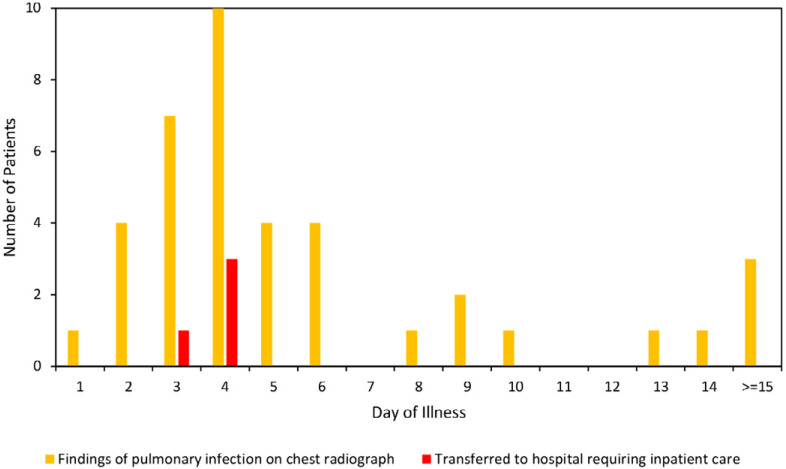
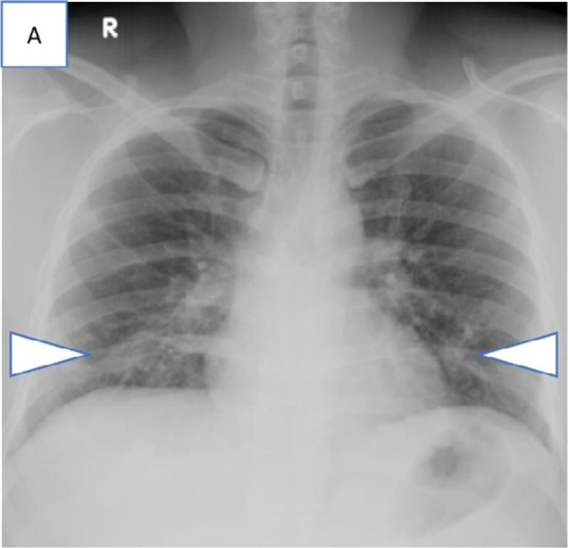
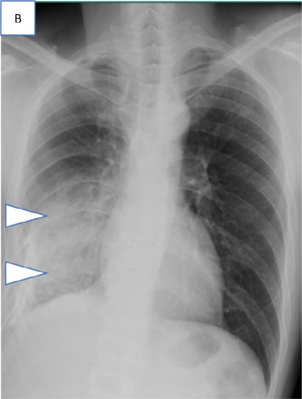
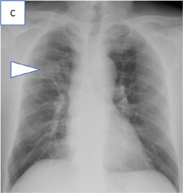
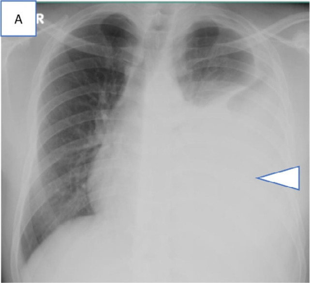
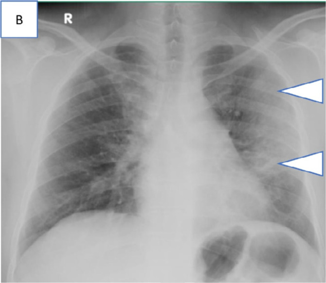
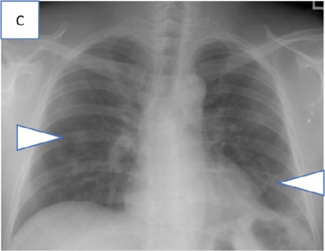
Background Singapore saw an escalation of coronavirus disease 2019 (COVID-19) cases from fewer than 4000 in April 2020 to more than 40 000 in June 2020, with most of these cases attributed to spread within shared facilities housing foreign workers. Appropriate triage and escalation of clinical care are crucial for this patient group managed in community care facilities (CCFs). Purpose To evaluate the imaging guideline recommendations for COVID-19 from the Fleischner Society and to analyze the clinical utility of screening chest radiography for asymptomatic or minimally symptomatic patients with COVID-19. Materials and Methods In this retrospective study, patients with reverse-transcription polymerase chain reaction-confirmed COVID-19 who were admitted to a designated CCF for continuation of their treatment during May 3-31, 2020, were identified. Upon admission, patients aged 36 years and older without any baseline chest images underwent chest radiography. All chest radiographs and clinical outcomes of patients, including those who were subsequently transferred to acute hospitals for escalation of care, were reviewed. Key proportions of patients with findings of pulmonary infection and those requiring further inpatient treatment were calculated, and 95% binomial proportion CIs were obtained using the Clopper-Pearson method. Results The study included 5621 patients. All patients were men (100%; 5621 of 5621), and the mean patient age was 37 years ± 8 (range, 17-60 years). A total of 1964 chest radiographs were obtained, of which normal images accounted for 98.0% (1925 of 1964 radiographs) and findings of pulmonary infection represented 2.0% (39 of 1964 radiographs). Only 0.2% of patients (four of 1964) with findings of pulmonary infection at chest radiography (all of whom were symptomatic) required supplemental oxygenation and inpatient treatment. None of the asymptomatic patients with findings of pulmonary infection required supplemental oxygenation, and they received only symptomatic treatment. Conclusion In accordance with Fleischner Society recommendations, screening chest radiography is not indicated in patients with coronavirus disease 2019 who are aged 17-60 years with mild or no symptoms unless there is risk of clinical deterioration. © RSNA, 2021 See also the editorial by Schaefer-Prokop and Prokop in this issue.


Figures

None
Graphical abstract
Workflow diagram illustrating the sequential stations of the admission process for patients with COVID-19 admitted into a community care facility, Singapore Expo, during May 3-31 for continuation of their management.
Figure 1:
Workflow diagram illustrating the sequential stations of the admission process for patients with COVID-19 admitted into a community care facility, Singapore Expo, during May 3-31 for continuation of their management.
Diagram illustrating the screening chest radiograph workflow for patients with COVID-19 admitted to a community care facility, Singapore Expo, during May 3-31. Patients aged 36 and older who had no baseline chest imaging received a screening chest radiograph. Additionally, patients who reported ill with respiratory symptoms during admission or subsequently to sick bay also received a chest radiograph regardless of age. Those with pulmonic changes on chest radiographs are either transferred to hospital if they are within 7 days of illness or deemed at risk of clinical deterioration.
Figure 2:
Diagram illustrating the screening chest radiograph workflow for patients with COVID-19 admitted to a community care facility, Singapore Expo, during May 3-31. Patients aged 36 and older who had no baseline chest imaging received a screening chest radiograph. Additionally, patients who reported ill with respiratory symptoms during admission or subsequently to sick bay also received a chest radiograph regardless of age. Those with pulmonic changes on chest radiographs are either transferred to hospital if they are within 7 days of illness or deemed at risk of clinical deterioration.
Screening chest radiograph findings of all patients with COVID-19 admitted to community care facility, Singapore Expo, by day of illness during May 3-31, 2020. Majority of patients (a) admitted to Singapore Expo were in the early phases of disease. Of the patients with screening chest radiograph, normal chest radiograph findings were most common throughout the disease course. Chest radiograph showing findings of pulmonary infection (b) showed a similar frequency trend in relation to symptom onset date. However, all hospitalizations requiring supplemental oxygenation or IV medication occurred in the first 4 days after symptom onset.
Figure 3a:
Screening chest radiograph findings of all patients with COVID-19 admitted to community care facility, Singapore Expo, by day of illness during May 3-31, 2020. Majority of patients (a) admitted to Singapore Expo were in the early phases of disease. Of the patients with screening chest radiograph, normal chest radiograph findings were most common throughout the disease course. Chest radiograph showing findings of pulmonary infection (b) showed a similar frequency trend in relation to symptom onset date. However, all hospitalizations requiring supplemental oxygenation or IV medication occurred in the first 4 days after symptom onset.
Figure 3b:
Screening chest radiograph findings of all patients with COVID-19 admitted to community care facility, Singapore Expo, by day of illness during May 3-31, 2020. Majority of patients (a) admitted to Singapore Expo were in the early phases of disease. Of the patients with screening chest radiograph, normal chest radiograph findings were most common throughout the disease course. Chest radiograph showing findings of pulmonary infection (b) showed a similar frequency trend in relation to symptom onset date. However, all hospitalizations requiring supplemental oxygenation or IV medication occurred in the first 4 days after symptom onset.
Series of chest radiograph in patients with COVID-19 pneumonia who were clinically asymptomatic and transferred out of the community care facility, Singapore Expo, to acute hospitals in view of findings of pulmonary infection on chest radiographs. Admission chest radiographs, A, in 36-year-old man on day 6 of illness showing bilateral lower zone patchy GGOs suspicious for infective foci (arrowheads), B, in 28-year-old man on day 2 of illness showing extensive consolidation (arrowheads) in the right mid and lower zone, and, C, in 55-year-old man on day 24 of illness with consolidation in the right upper zone (arrowhead).
Figure 4a:
Series of chest radiograph in patients with COVID-19 pneumonia who were clinically asymptomatic and transferred out of the community care facility, Singapore Expo, to acute hospitals in view of findings of pulmonary infection on chest radiographs. Admission chest radiographs, A, in 36-year-old man on day 6 of illness showing bilateral lower zone patchy GGOs suspicious for infective foci (arrowheads), B, in 28-year-old man on day 2 of illness showing extensive consolidation (arrowheads) in the right mid and lower zone, and, C, in 55-year-old man on day 24 of illness with consolidation in the right upper zone (arrowhead).
Figure 4b:
Series of chest radiograph in patients with COVID-19 pneumonia who were clinically asymptomatic and transferred out of the community care facility, Singapore Expo, to acute hospitals in view of findings of pulmonary infection on chest radiographs. Admission chest radiographs, A, in 36-year-old man on day 6 of illness showing bilateral lower zone patchy GGOs suspicious for infective foci (arrowheads), B, in 28-year-old man on day 2 of illness showing extensive consolidation (arrowheads) in the right mid and lower zone, and, C, in 55-year-old man on day 24 of illness with consolidation in the right upper zone (arrowhead).
Figure 4c:
Series of chest radiograph in patients with COVID-19 pneumonia who were clinically asymptomatic and transferred out of the community care facility, Singapore Expo, to acute hospitals in view of findings of pulmonary infection on chest radiographs. Admission chest radiographs, A, in 36-year-old man on day 6 of illness showing bilateral lower zone patchy GGOs suspicious for infective foci (arrowheads), B, in 28-year-old man on day 2 of illness showing extensive consolidation (arrowheads) in the right mid and lower zone, and, C, in 55-year-old man on day 24 of illness with consolidation in the right upper zone (arrowhead).
Series of chest radiograph in patients with COVID-19 pneumonia who were transferred out of community care facility, Singapore Expo, to acute hospitals in view of respiratory symptoms or deranged vitals and subsequently required supplemental oxygen. Admission chest radiographs, A, in a 41-year-old man on day 3 of illness with a large left pleural effusion and adjacent consolidation (arrowhead) treated for concomitant pleural tuberculosis, B, in a 40-year-old man on day 4 of illness with consolidation across the left mid and upper zones (arrowheads), and, C, in a 42-year-old man on day 4 of illness with patchy GGOs in the peripheral aspects of both the mid zone and the left lower zone (arrowheads).
Figure 5a:
Series of chest radiograph in patients with COVID-19 pneumonia who were transferred out of community care facility, Singapore Expo, to acute hospitals in view of respiratory symptoms or deranged vitals and subsequently required supplemental oxygen. Admission chest radiographs, A, in a 41-year-old man on day 3 of illness with a large left pleural effusion and adjacent consolidation (arrowhead) treated for concomitant pleural tuberculosis, B, in a 40-year-old man on day 4 of illness with consolidation across the left mid and upper zones (arrowheads), and, C, in a 42-year-old man on day 4 of illness with patchy GGOs in the peripheral aspects of both the mid zone and the left lower zone (arrowheads).
Figure 5b:
Series of chest radiograph in patients with COVID-19 pneumonia who were transferred out of community care facility, Singapore Expo, to acute hospitals in view of respiratory symptoms or deranged vitals and subsequently required supplemental oxygen. Admission chest radiographs, A, in a 41-year-old man on day 3 of illness with a large left pleural effusion and adjacent consolidation (arrowhead) treated for concomitant pleural tuberculosis, B, in a 40-year-old man on day 4 of illness with consolidation across the left mid and upper zones (arrowheads), and, C, in a 42-year-old man on day 4 of illness with patchy GGOs in the peripheral aspects of both the mid zone and the left lower zone (arrowheads).
Figure 5c:
Series of chest radiograph in patients with COVID-19 pneumonia who were transferred out of community care facility, Singapore Expo, to acute hospitals in view of respiratory symptoms or deranged vitals and subsequently required supplemental oxygen. Admission chest radiographs, A, in a 41-year-old man on day 3 of illness with a large left pleural effusion and adjacent consolidation (arrowhead) treated for concomitant pleural tuberculosis, B, in a 40-year-old man on day 4 of illness with consolidation across the left mid and upper zones (arrowheads), and, C, in a 42-year-old man on day 4 of illness with patchy GGOs in the peripheral aspects of both the mid zone and the left lower zone (arrowheads).

Comment in

References

    1. Li Q, Guan X, Wu P, Wang X, Zhou L, Tong Y, et al. Early transmission dynamics in Wuhan, China, of novel coronavirus-infected pneumonia. N Engl J Med. 2020;382(13):1199–207. - PMC - PubMed
    1. World Health Organization. Coronavirus Disease (COVID-19) [Internet]. 2020 [cited 2020 Jun 17]. Available from: https://www.who.int/emergencies/diseases/novel-coronavirus-2019
    1. Nicola M, Alsafi Z, Sohrabi C, Kerwan A, Al-Jabir A, Iosifidis C, et al. The socio-economic implications of the coronavirus pandemic (COVID-19): A review [Internet]. Vol. 78, International Journal of Surgery. IJS Publishing Group Ltd; 2020. 185–193 p. Available from: 10.1016/j.ijsu.2020.04.018 - DOI - PMC - PubMed
    1. Dong E, Du H, Gardner L. An interactive web-based dashboard to track COVID-19 in real time. Lancet Infect Dis [Internet]. 2020;20(5):533–4. Available from: 10.1016/S1473-3099(20)30120-1 - DOI - PMC - PubMed
    1. Lee VJ, Chiew CJ, Khong WX. Interrupting transmission of COVID-19: lessons from containment efforts in Singapore. J Travel Med. 2020 May;27(3). - PMC - PubMed

LinkOut - more resources