Review
. 2021 Apr;299(1):E193-E203.
doi: 10.1148/radiol.2020204267. Epub 2020 Dec 8.

RSNA International Trends: A Global Perspective on the COVID-19 Pandemic and Radiology in Late 2020

Affiliations
Review

RSNA International Trends: A Global Perspective on the COVID-19 Pandemic and Radiology in Late 2020

Bien Soo Tan et al. Radiology. 2021 Apr.

Abstract

The coronavirus disease 2019 pandemic has challenged and changed health care systems around the world. There has been a heterogeneity of disease burden, health care resources, and nonimaging testing availability, both geographically and over time. In parallel, there has been a continued increase in understanding how the disease affects patients, effectiveness of therapeutic options, and factors that modulate transmission risk. In this report, radiology experts in representative countries from around the world share insights gained from local experience. These insights provide a guidepost to help address management challenges as cases continue to rise in many parts of the world and suggest modifications in workflow that are likely to continue after this pandemic subsides.


Figures

COVID-19 cases over time by continent. The world’s population is severely affected by this pandemic, with many regions showing increasing cases in the later part of 2020. While healthcare systems and radiology practice have rapidly adapted best practices learned from initial experiences, the growth in COVID-19 incidence will continue to test how best to manage the needs of patients. Data from World Health Organization (WHO), https://covid19.who.int/table, 2020/11/06.
Figure 1.
COVID-19 cases over time by continent. The world’s population is severely affected by this pandemic, with many regions showing increasing cases in the later part of 2020. While healthcare systems and radiology practice have rapidly adapted best practices learned from initial experiences, the growth in COVID-19 incidence will continue to test how best to manage the needs of patients. Data from World Health Organization (WHO), https://covid19.who.int/table, 2020/11/06.
Total COVID-19 cases to date in representative countries. The heterogeneity of disease burden is in part determined by national or regional mitigation strategies. These case incidences determine the COVID-related need and utilization of overall health care and radiology resources in different countries. Data from World Bank, https://data.worldbank.org/indicator/SP.POP.TOTL, 2019 data and Johns Hopkins Dashboard, https://coronavirus.jhu.edu/map.html, 10/27/2020.
Figure 2.
Total COVID-19 cases to date in representative countries. The heterogeneity of disease burden is in part determined by national or regional mitigation strategies. These case incidences determine the COVID-related need and utilization of overall health care and radiology resources in different countries. Data from World Bank, https://data.worldbank.org/indicator/SP.POP.TOTL, 2019 data and Johns Hopkins Dashboard, https://coronavirus.jhu.edu/map.html, 10/27/2020.
Highlights of RSNA’s efforts. The rapid dissemination of the latest COVID-19 research, education on imaging interpretation, and workflow best practices is vital to the health of patients, both for diagnosis and treatment assessment. The insights allow improved workflows to increase the safety of patients and healthcare workers. The broad RSNA connectivity throughout the imaging community ensures radiologists globally remain fully up to date. Data source: RSNA internal data/www.rsna.org
Figure 3.
Highlights of RSNA’s efforts. The rapid dissemination of the latest COVID-19 research, education on imaging interpretation, and workflow best practices is vital to the health of patients, both for diagnosis and treatment assessment. The insights allow improved workflows to increase the safety of patients and healthcare workers. The broad RSNA connectivity throughout the imaging community ensures radiologists globally remain fully up to date. Data source: RSNA internal data/www.rsna.org
COVID-19 Chest AI workflow in Radvid19 demonstrates academic, private, and government partnership created in Brazil. This workflow is free to users in Brazil and allowed high level automatic interpretation and quantitative lung involvement results even to remote hospitals in 10 min, helping address the increased demand for COVID-19 related image interpretation in rural parts of the country.
Figure 4:
COVID-19 Chest AI workflow in Radvid19 demonstrates academic, private, and government partnership created in Brazil. This workflow is free to users in Brazil and allowed high level automatic interpretation and quantitative lung involvement results even to remote hospitals in 10 min, helping address the increased demand for COVID-19 related image interpretation in rural parts of the country.
The Brazilian COVID-19 Chest CT Database. There are 47 hospitals connected from different Brazilian states to the central database. Almost 20,000 chest CT’s have been analyzed to date, with a COVID-19 prevalence of 75%. This high prevalence is in part a reflection of increased cases submitted for analysis from dedicated COVID-19 and reference hospitals. The results screen shows example images, the probability of COVID-19 and the percent of lung parenchyma involvement. Such automated analysis can aid in direct patient management, as well as be potentially utilized for studies evaluating the efficacy of therapeutic interventions.
Figure 5:
The Brazilian COVID-19 Chest CT Database. There are 47 hospitals connected from different Brazilian states to the central database. Almost 20,000 chest CT’s have been analyzed to date, with a COVID-19 prevalence of 75%. This high prevalence is in part a reflection of increased cases submitted for analysis from dedicated COVID-19 and reference hospitals. The results screen shows example images, the probability of COVID-19 and the percent of lung parenchyma involvement. Such automated analysis can aid in direct patient management, as well as be potentially utilized for studies evaluating the efficacy of therapeutic interventions.
COVID-19 Virtual Assistant.Effective integration of AI into new radiology workflows help efficient patient communication and educationwith the use of a chatbot. Virtual assistants can evaluate risk self-assessment including the need for immediate attention, provide access to speak to a provider, help schedule a visit, and provide up-to-date information about COVID-19 testing sites and availability, visitor policies, and other patient care information.
Figure 6:
COVID-19 Virtual Assistant. Effective integration of AI into new radiology workflows help efficient patient communication and educationwith the use of a chatbot. Virtual assistants can evaluate risk self-assessment including the need for immediate attention, provide access to speak to a provider, help schedule a visit, and provide up-to-date information about COVID-19 testing sites and availability, visitor policies, and other patient care information.
Preparing for a portable interventional radiology procedure in a COVID-19 isolation room.Full PPE with N-95 respirator and face shield. The level of PPE should be concordant with risk of aerosolized particles generated during a procedure.
Figure 7:
Preparing for a portable interventional radiology procedure in a COVID-19 isolation room. Full PPE with N-95 respirator and face shield. The level of PPE should be concordant with risk of aerosolized particles generated during a procedure.

References

    1. RSNA News article: https://www.rsna.org/news/2020/June/COVID-Open-Radiology-Database.
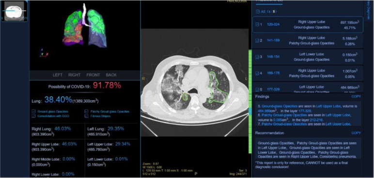
    1. Wong HYF, Lam HYS, Fong AH, Leung ST, Chin TW, Lo CSY, Lui MM, Lee JCY, Chiu KW, Chung TW, Lee EYP, Wan EYF, Hung IFN, Lam TPW, Kuo MD, Ng MY. Frequency and Distribution of Chest Radiographic Findings in Patients Positive for COVID-19. Radiology 2020;296(2):E72-e78. doi: 10.1148/radiol.2020201160. - PMC - PubMed
    1. Fang Y, Zhang H, Xu Y, Xie J, Pang P, Ji W. CT Manifestations of Two Cases of 2019 Novel Coronavirus (2019-nCoV) Pneumonia. Radiology 2020;295(1):208-209. doi: 10.1148/radiol.2020200280. - PMC - PubMed
    1. Song F, Shi N, Shan F, Zhang Z, Shen J, Lu H, Ling Y, Jiang Y, Shi Y. Emerging 2019 Novel Coronavirus (2019-nCoV) Pneumonia. Radiology 2020;295(1):210-217. doi: 10.1148/radiol.2020200274. - PMC - PubMed
    1. Li M, Lei P, Zeng H, Li Z, Yu P, Fan B, Wang C, Li Z, Zhou Z, Hu S. and Liu H, Coronavirus Disease (COVID-19): Spectrum of CT Findings and Temporal Progression of the Disease. Acad Radiol 2020; 27:603-608. - PMC - PubMed

LinkOut - more resources