

Predictive value of CT imaging findings in COVID-19 pneumonia at the time of first-screen regarding the need for hospitalization or intensive care unit
- PMID: 33290242
- PMCID: PMC8480949
- DOI: 10.5152/dir.2020.20421
Predictive value of CT imaging findings in COVID-19 pneumonia at the time of first-screen regarding the need for hospitalization or intensive care unit
Abstract
Purpose: In this study, we aimed to reveal the relationship between initial lung parenchymal involvement patterns and the subsequent need for hospitalization and/or intensive care unit admission in coronavirus disease 2019 (COVID-19) positive cases.
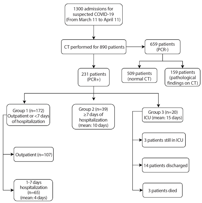
Methods: Overall, 231 patients diagnosed with COVID-19 as proven by PCR were included in this study. Based on the duration of hospitalization, patients were divided into three groups as follows: Group 1, patients receiving outpatient treatment or requiring hospitalization <7 days; Group 2, requiring hospitalization ≥7 days; Group 3, patients requiring at least 1 day of intensive care at any time. Chest CT findings at first admission were evaluated for the following features: typical/atypical involvement of the disease, infiltration patterns (ground-glass opacities, crazy-paving pattern, consolidation), distribution and the largest diameters of the lesions, total lesion numbers, number of affected lung lobes, and affected total lung parenchyma percentages. The variability of all these findings according to the groups was analyzed statistically.
Results: In this study, 172 patients were in Group 1, 39 patients in Group 2, and 20 patients in Group 3. The findings obtained in this study indicated that there was no statistically significant difference in ground-glass opacity rates among the groups (p = 0.344). The rates of crazy-paving and consolidation patterns were significantly higher in Groups 2 and 3 than in Group 1 (p = 0.001, p = 0.002, respectively). The rate of right upper, left upper lobe, and right middle lobe involvements as consolidation pattern was significantly higher in Group 3 than in Group 1 (p = 0.148, p = 0.935, p = 0.143, respectively). A statistically significant difference was also found between the affected lobe numbers, total lesion numbers, the diameter of the largest lesion, and the affected lung parenchyma percentages between the groups (p = 0.001). The average number of impacted lobes in Group 1 was 2; 4 in Group 2 and Group 3. The mean percentage of affected lung parenchyma percentage was 25% in Group 1 and Group 2, and 50% in Group 3.
Conclusion: In case of infiltration dominated by right middle or upper lobe involvement with a consolidation pattern, there is a higher risk of future intensive care need. Also, the need for intensive care increases as the number of affected lobes and percentage of affected parenchymal involvement increase.
Conflict of interest statement
The authors declared no conflicts of interest.
Figures
Similar articles
-
Prognostic findings for ICU admission in patients with COVID-19 pneumonia: baseline and follow-up chest CT and the added value of artificial intelligence.Emerg Radiol. 2022 Apr;29(2):243-262. doi: 10.1007/s10140-021-02008-y. Epub 2022 Jan 20. Emerg Radiol. 2022. PMID: 35048222 Free PMC article. Review.
-
[Spatial and temporal distribution and predictive value of chest CT scoring in patients with COVID-19].Zhonghua Jie He He Hu Xi Za Zhi. 2021 Mar 12;44(3):230-236. doi: 10.3760/cma.j.cn112147-20200522-00626. Zhonghua Jie He He Hu Xi Za Zhi. 2021. PMID: 33721937 Chinese.
-
CT imaging features of COVID-19 pneumonia: initial experience from Turkey.Diagn Interv Radiol. 2020 Jul;26(4):308-314. doi: 10.5152/dir.2020.20307. Diagn Interv Radiol. 2020. PMID: 32558645 Free PMC article.
-
Temporal relationship between serial RT-PCR results and serial chest CT imaging, and serial CT changes in coronavirus 2019 (COVID-19) pneumonia: a descriptive study of 155 cases in China.Eur Radiol. 2021 Mar;31(3):1175-1184. doi: 10.1007/s00330-020-07268-9. Epub 2020 Sep 15. Eur Radiol. 2021. PMID: 32930834 Free PMC article.
-
Coronavirus Disease 2019 (COVID-19): A Systematic Review of Imaging Findings in 919 Patients.AJR Am J Roentgenol. 2020 Jul;215(1):87-93. doi: 10.2214/AJR.20.23034. Epub 2020 Mar 14. AJR Am J Roentgenol. 2020. PMID: 32174129
Cited by
-
Predicting Severe Respiratory Failure in Patients with COVID-19: A Machine Learning Approach.J Clin Med. 2024 Dec 4;13(23):7386. doi: 10.3390/jcm13237386. J Clin Med. 2024. PMID: 39685844 Free PMC article.
-
Detection of COVID-19 and its pulmonary stage using Bayesian hyperparameter optimization and deep feature selection methods.Expert Syst. 2022 Sep 26:e13141. doi: 10.1111/exsy.13141. Online ahead of print. Expert Syst. 2022. PMID: 36245832 Free PMC article.
-
Effectiveness of Tocilizumab in Patients with Severe or Critical Lung Involvement in COVID-19: A Retrospective Study.J Clin Med. 2022 Apr 20;11(9):2286. doi: 10.3390/jcm11092286. J Clin Med. 2022. PMID: 35566412 Free PMC article.
-
Time-dependent CT score-based model for identifying severe/critical COVID-19 at a fever clinic after the emergence of Omicron variant.Heliyon. 2024 Mar 25;10(7):e27963. doi: 10.1016/j.heliyon.2024.e27963. eCollection 2024 Apr 15. Heliyon. 2024. PMID: 38586383 Free PMC article.
-
Prognostic findings for ICU admission in patients with COVID-19 pneumonia: baseline and follow-up chest CT and the added value of artificial intelligence.Emerg Radiol. 2022 Apr;29(2):243-262. doi: 10.1007/s10140-021-02008-y. Epub 2022 Jan 20. Emerg Radiol. 2022. PMID: 35048222 Free PMC article. Review.
References
MeSH terms
LinkOut - more resources
Full Text Sources
Medical
