

Angiography with optical coherence tomography as a biomarker in multiple sclerosis
- PMID: 33290417
- PMCID: PMC7723290
- DOI: 10.1371/journal.pone.0243236
Angiography with optical coherence tomography as a biomarker in multiple sclerosis
Abstract
Purpose: To investigate superficial retinal microvascular plexuses detected by optical coherence tomography angiography (OCT-A) in multiple sclerosis (MS) subjects and compare them with healthy controls.
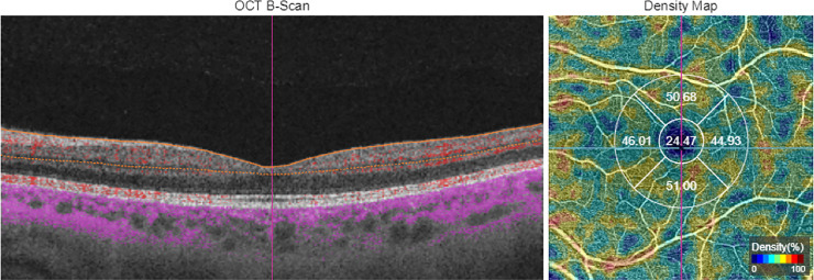
Methods: A total of 92 eyes from 92 patients with relapsing-remitting MS and 149 control eyes were included in this prospective observational study. OCT-A imaging was performed using Triton Swept-Source OCT (Topcon Corporation, Japan). The vessel density (VD) percentage in the superficial retinal plexus and optic disc area (6 x 6 mm grid) was measured and compared between groups.
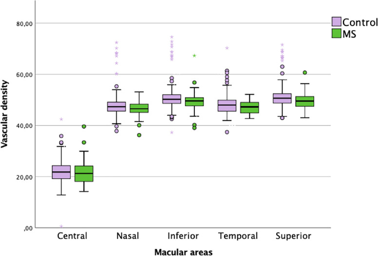
Results: MS patients showed a significant decrease VD in the superior (p = 0.005), nasal (p = 0.029) and inferior (p = 0.040) parafoveal retina compared with healthy subjects. Patients with disease durations of more than 5 years presented lower VD in the superior (p = 0.002), nasal (p = 0.017) and inferior (p = 0.022) parafoveal areas compared with healthy subjects. Patients with past optic neuritis episodes did not show retinal microvasculature alterations, but patients with an EDSS score of less than 3 showed a significant decrease in nasal (p = 0.024) and superior (p = 0.006) perifoveal VD when compared with healthy subjects.
Conclusions: MS produces a decrease in retinal vascularization density in the superficial plexus of the parafoveal retina. Alterations in retinal vascularization observed in MS patients are independent of the presence of optic nerve inflammation. OCT-A has the ability to detect subclinical vascular changes and is a potential biomarker for diagnosing the presence and progression of MS.
Conflict of interest statement
The authors have declared that no competing interests exist.
Figures
Similar articles
-
OCT and OCT-A biomarkers in multiple sclerosis - review.Rom J Ophthalmol. 2023 Apr-Jun;67(2):107-110. doi: 10.22336/rjo.2023.20. Rom J Ophthalmol. 2023. PMID: 37522023 Free PMC article. Review.
-
Optical Coherence Tomography Angiography of Optic Disc and Macula Vessel Density in Glaucoma and Healthy Eyes.J Glaucoma. 2019 Jan;28(1):80-87. doi: 10.1097/IJG.0000000000001125. J Glaucoma. 2019. PMID: 30461553
-
Peripapillary and parafoveal microvascular changes in eyes with optic neuritis and their fellow eyes measured by optical coherence tomography angiography: an Exploratory Study.Acta Ophthalmol. 2021 May;99(3):288-298. doi: 10.1111/aos.14577. Epub 2020 Aug 24. Acta Ophthalmol. 2021. PMID: 32833336
-
Optical coherence tomography angiography indicates associations of the retinal vascular network and disease activity in multiple sclerosis.Mult Scler. 2019 Feb;25(2):224-234. doi: 10.1177/1352458517750009. Epub 2018 Jan 5. Mult Scler. 2019. PMID: 29303033
-
Microvascular impairments detected by optical coherence tomography angiography in multiple sclerosis patients: A systematic review and meta-analysis.Front Neurosci. 2023 Jan 13;16:1121899. doi: 10.3389/fnins.2022.1121899. eCollection 2022. Front Neurosci. 2023. PMID: 36711144 Free PMC article.
Cited by
-
The Heterogeneous Multiple Sclerosis Lesion: How Can We Assess and Modify a Degenerating Lesion?Int J Mol Sci. 2023 Jul 5;24(13):11112. doi: 10.3390/ijms241311112. Int J Mol Sci. 2023. PMID: 37446290 Free PMC article. Review.
-
Association of the retinal vasculature, intrathecal immunity, and disability in multiple sclerosis.Front Immunol. 2022 Nov 11;13:997043. doi: 10.3389/fimmu.2022.997043. eCollection 2022. Front Immunol. 2022. PMID: 36439131 Free PMC article.
-
OCT and OCT-A biomarkers in multiple sclerosis - review.Rom J Ophthalmol. 2023 Apr-Jun;67(2):107-110. doi: 10.22336/rjo.2023.20. Rom J Ophthalmol. 2023. PMID: 37522023 Free PMC article. Review.
-
New Method of Early RRMS Diagnosis Using OCT-Assessed Structural Retinal Data and Explainable Artificial Intelligence.Transl Vis Sci Technol. 2025 Feb 3;14(2):14. doi: 10.1167/tvst.14.2.14. Transl Vis Sci Technol. 2025. PMID: 39928305 Free PMC article.
-
Barriers in Healthcare to the Use of Optical Coherence Tomography Angiography in Multiple Sclerosis.Neurol Ther. 2025 Feb;14(1):45-56. doi: 10.1007/s40120-024-00670-1. Epub 2024 Nov 5. Neurol Ther. 2025. PMID: 39500829 Free PMC article. Review.
References
-
- Noseworthy JH, Lucchinetti C, Rodriguez M, Weinshenker BG. Multiple Sclerosis. N Engl J Med.2000;343: 938–52. - PubMed
-
- Prineas J. Pathology of the early lesion in multiple sclerosis. Hum Pathol: 1975; 6(5): 531–554. - PubMed
-
- Gordon-Lipkin E, Chodkowski B, Reich D. S, Smith S. A, Pulicken M, Balcer L. J, et al. Retinal nerve fiber layer is associated with brain atrophy in multiple sclerosis, Neurology. 2007;69(16): 1603–1609 - PubMed
MeSH terms
LinkOut - more resources
Full Text Sources
Medical
Miscellaneous
