. 2021 Jan 11;49(1):114-126.
doi: 10.1093/nar/gkaa1130.

Histone benzoylation serves as an epigenetic mark for DPF and YEATS family proteins

Affiliations

Histone benzoylation serves as an epigenetic mark for DPF and YEATS family proteins

Xiangle Ren et al. Nucleic Acids Res. .

Abstract

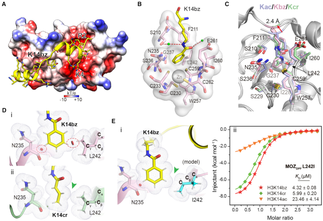
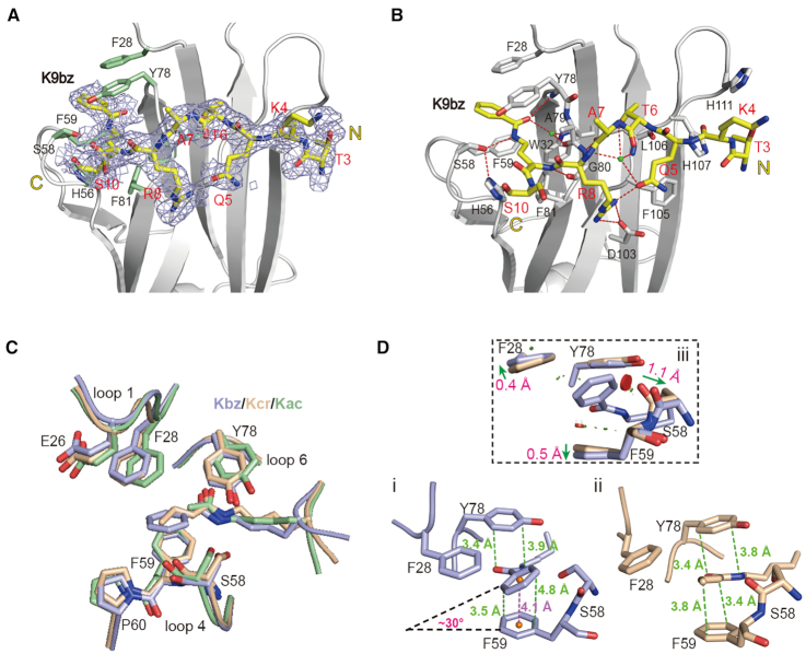
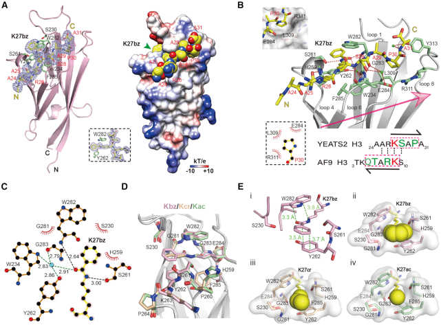
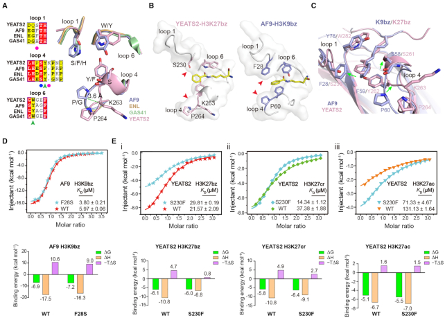
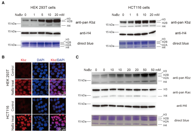
Histone modifications and their functional readout serve as an important mechanism for gene regulation. Lysine benzoylation (Kbz) on histones is a recently identified acylation mark associated with active transcription. However, it remains to be explored whether putative readers exist to recognize this epigenetic mark. Here, our systematic binding studies demonstrated that the DPF and YEATS, but not the Bromodomain family members, are readers for histone Kbz. Co-crystal structural analyses revealed a 'hydrophobic encapsulation' and a 'tip-sensor' mechanism for Kbz readout by DPF and YEATS, respectively. Moreover, the DPF and YEATS family members display subtle yet unique features to create somewhat flexible engagements of different acylation marks. For instance, YEATS2 but not the other YEATS proteins exhibits best preference for Kbz than lysine acetylation and crotonylation due to its wider 'tip-sensor' pocket. The levels of histone benzoylation in cultured cells or in mice are upregulated upon sodium benzoate treatment, highlighting its dynamic regulation. In summary, our work identifies the first readers for histone Kbz and reveals the molecular basis underlying Kbz recognition, thus paving the way for further functional dissections of histone benzoylation.


Figures

Figure 1.
Figure 1.
Identification of DPF and YEATS domains as readers for histone benzoylation. (A) Chemical formula of twelve histone lysine acylations. The acylation group is color shaded and benzoylation is highlighted in bold. (B) ITC fitting curves of indicated histone peptides with human DPF domains. Mean KD and standard deviation are shown (N ≥ 3). Un, unmodified peptide. (C) Affinity comparison of Kbz with Kcr or Kac among DPF family members. Red dashes, reference line of equal affinity; Blue dashes, reference line of 1:4 preference. (D) ITC fitting curves of indicated histone peptides with human YEATS domains. Mean KD and standard deviation are shown (N ≥ 3). N.D., not detectable. (E) Affinity comparison of Kbz with Kcr or Kac among YEATS family members. Red dashes, reference line of equal affinity.
Figure 2.
Figure 2.
Molecular details for histone H3K14bz recognition by MOZDPF. (A) Overall structure of MOZDPF in complex with H31–25K14bz. MOZDPF is shown as electrostatic potential surface ranging from −10 (red) to +10 (blue) kT/e. Histone H3 is shown as yellow cartoon with residues depicted as sticks. (B) Recognition of K14bz mark by the hydrophobic reader pocket of MOZDPF. Small green balls, water molecules; Red dashes, hydrogen bonds; Gray sphere, zinc ion. (C) Superimposition of K14bz-bound (pink), K14ac-bound (light blue, PDB: 4LLB) and K14cr-bound (pale green, PDB ID: 5B76) MOZDPF structures. Key pocket residues are depicted as sticks; Red arrow highlights the up-lift of the H3K14bz backbone. (D) Close contact analyses of MOZDPF structure bound to (i) H3K14bz and (ii) H3K14cr (PDB ID: 5B76). Gray dots denote van der Waals surface of the indicated residues. Large red disk, severe van der Waals overlap; Small green disk, slight van der Waals overlap. Red circles and green arrowhead highlight steric clashes in H3K14bz imbedded structure and spatial compatibility in H3K14cr imbedded structure, respectively. Hydrogens (white sticks) were added for analysis. (E) i, structural modeling of L242I mutation of MOZDPF bound to H3K14bz. Note the loss of ‘close contact’ in L242I mutant (green arrowhead); ii, ITC fitting curves of the indicated histone peptides with L242I mutant. Mean KD and standard deviation are shown (N = 2).
Figure 3.
Figure 3.
Molecular recognition of histone benzoylation by AF9YEATS. (A) Fo-Fc omit map of H3K9bz peptide contoured at 2.0 σ level. Histone peptide is shown as yellow sticks. Key residues of AF9YEATS are depicted as pale green sticks. Light blue meshes, Fo-Fc omit map. (B) Hydrogen bonding networks between H3K9bz peptide and AF9YEATS. (C) Structure alignment of K9bz-bound (light blue), K9ac-bound (pale green, PDB ID: 4TMP) and K9cr-bound (wheat, PDB ID: 5HJB) structures. (D) π–π stacking analyses of (i) K9bz and (ii) K9cr in AF9YEATS reader pocket and its local conformational adjustments (iii). Distances measured between atoms or centroids are color-coded in green or magenta, respectively. Symbols of close contacts are as described for Figure 2D. Green arrows, conformational displacement comparing Kbz and Kcr readout.
Figure 4.
Figure 4.
Structural basis for Kbz recognition by YEATS2YEATS. (A) Overall structure of YEATS2YEATS bound to H3K27bz peptide. Left, ribbon view; Right, electrostatic potential surface view. H3K27bz peptide was covered by Fo-Fc omit map contoured at 2.0 σ level. (B) Binding details between YEATS2YEATS and H3K27bz peptide. A sequence comparison of H3K27 and H3K9 is shown below. Note the positioning of P30 in a hydrophobic pocket of YEATS2YEATS. (C) LigPlot diagram listing critical contacts between K27bz and YEATS2YEATS. The schematic symbols are described in Supplementary Figure S2B. (D) Alignment of K27bz-bound (light pink), K27ac-bound (pale green, PDB ID: 5XNV) and K27cr-bound (wheat, PDB ID: 5IQL) structures. (E) i, π–π stacking analysis of K27bz bound to YEATS2YEATS; Positioning of (ii) Kbz, (iii) Kcr and (iv) Kac into the YEATS2YEATS reader pocket. Green dashes, π–π distances; Yellow spheres, different acyl groups.
Figure 5.
Figure 5.
A ‘tip-sensor’ mechanism determines acylation type selectivity of YEATS domains. (A) Sequence and structural alignments of reader pocket loops among four human YEATS domains. Magenta circles, ‘tip-sensor’ residues; Blue hexagon, conserved Ser residue for acyllysine amide recognition; Green arrowhead, aromatic sandwiching residues; Light blue, AF9YEATS; Wheat, ENLYEATS (PDB ID: 5J9S); Pale green, GAS41YEATS (PDB: 5XTZ); Light pink, YEATS2YEATS. (B) Comparison of reader pocket between YEATS2YEATS and AF9YEATS. Red arrowheads indicate a wide and narrow end-opening of YEATS2 and AF9, respectively. (C) Structure alignment of YEATS2YEATS-H3K27bz with AF9YEATS-H3K9bz centred on the recognition pocket. Green arrows indicate conformational variations between the two proteins. (D) ITC assays of Kbz binding by WT AF9YEATS and its F28S mutant. Mean KD and standard deviation are shown (N = 4). Thermodynamic parameters of each titration are illustrated below. (E) ITC assays of (i) Kbz, (ii) Kcr and (iii) Kac binding by WT YEATS2YEATS and its S230F mutant. Mean KD and standard deviation are shown (N ≥ 2). Thermodynamic parameters of each titration are illustrated below.
Figure 6.
Figure 6.
Histone benzoylation is an inducible epigenetic mark in cells and mice. (A) Western blot analysis of core histone Kbz levels in response to the indicated concentrations of NaBz treatment in HEK 293T and HCT116 cells. Unmodified histone H4 was used as a loading control. (B) Immunofluorescence analysis of Kbz levels in HEK 293T and HCT116 cells. DNA was stained with DAPI. Control, cells without NaBz treatment. (C) Western blot analysis of core histone Kbz and Kac levels in mouse intestinal epithelial cells after the indicated concentrations of NaBz water feeding. Unmodified histone H4 was used as a loading control. Assays were performed in duplicates by using two mice for each concentration.

References

    1. Strahl B.D., Allis C.D.. The language of covalent histone modifications. Nature. 2000; 403:41–45. - PubMed
    1. Waddington C.H. The epigenotype. 1942. Int. J. Epidemiol. 2012; 41:10–13. - PubMed
    1. Phillips D.M. The presence of acetyl groups of histones. Biochem. J. 1963; 87:258–263. - PMC - PubMed
    1. Jiang T., Zhou X., Taghizadeh K., Dong M., Dedon P.C.. N-formylation of lysine in histone proteins as a secondary modification arising from oxidative DNA damage. Proc. Natl. Acad. Sci. U.S.A. 2007; 104:60–65. - PMC - PubMed
    1. Chen Y., Sprung R., Tang Y., Ball H., Sangras B., Kim S.C., Falck J.R., Peng J., Gu W., Zhao Y.. Lysine propionylation and butyrylation are novel post-translational modifications in histones. Mol. Cell. Proteomics. 2007; 6:812–819. - PMC - PubMed

Publication types

MeSH terms