Review
. 2021 Apr 16;22(8):1347-1356.
doi: 10.1002/cbic.202000745. Epub 2021 Feb 3.

Engineered Sortases in Peptide and Protein Chemistry

Affiliations
Review

Engineered Sortases in Peptide and Protein Chemistry

Christian Freund et al. Chembiochem. .

Abstract

The transpeptidase sortase A of Staphylococcus aureus (Sa-SrtA) is a valuable tool in protein chemistry. The native enzyme anchors surface proteins containing a highly conserved LPxTG sorting motif to a terminal glycine residue of the peptidoglycan layer in Gram-positive bacteria. This reaction is exploited for sortase-mediated ligation (SML), allowing the site-specific linkage of synthetic peptides and recombinant proteins by a native peptide bond. However, the moderate catalytic efficiency and specificity of Sa-SrtA fueled the development of new biocatalysts for SML, including the screening of sortase A variants form microorganisms other than S. aureus and the directed protein evolution of the Sa-SrtA enzyme itself. Novel display platforms and screening formats were developed to isolate sortases with altered properties from mutant libraries. This yielded sortases with strongly enhanced catalytic activity and enzymes recognizing new sorting motifs as substrates. This minireview focuses on recent advances in the field of directed sortase evolution and applications of these tailor-made enzymes in biochemistry.

Keywords: protein bioconjugation; protein engineering; protein semisynthesis; sortases; transpeptidases.


Conflict of interest statement

The authors declare no conflict of interest.

Figures
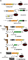
Figure 1
Figure 1
Sortases and sortase‐mediated ligation (SML). a) Sortase A of S. aureus (Sa‐SrtA) recognizes LPxTG sorting motifs as substrates and cleaves this sequence at the threonine residue with formation of a sortase‐bound thioester. The downstream peptide with N‐terminal glycine residue is released from the enzyme. The thioester is resolved by ligation to a peptide nucleophile with at least two glycine residues at the N terminus. b) Native sortases install surface proteins to the bacterial peptidoglycan cross‐bridge. In the case of S. aureus, the cross‐bridge contains a pentaglycine moiety as the nucleophile, while the peptidoglycan of S. pyogenes provides an N‐terminal alanine residue for the ligation reaction. The composition of the bacterial peptidoglycan impacts sortase substrate specificity. c) Sortase A of Corynebacterium diphtheriae catalyzes isopeptide formation at an internal lysine of SpaA with a C‐terminal sorting motif from a neighboring SpaA during pilin polylmerization. SP: surface protein.
Figure 2
Figure 2
Selection schemes for sortase A activity enhancement. a) General scheme for evolving bond‐forming enzymes by yeast display as applied to sortase A. SrtA of S. aureus is displayed as a fusion with the surface protein Aga2p (not shown), which itself is covalently linked to Agap1 (also not shown) carrying the reactive S6 peptide. Sfp phosphopantetheinyl transferase from Bacillus subtilis is then used to covalently link two glycine residues to the S6 tag, which provide the nucleophile for the sortase reaction. Biotinylated sorting motif peptide is then ligated to the N‐terminal glycine residue by the spatially proximal sortase A protein, thereby coupling genotype with phenotype. Streptavidin‐phycoerythrin is then used to detect and select successfully ligated product by FACS. b) a FRET‐based platform for srtA activity screening. The LPETG sorting motif was fused to the C terminus of EGFP, while a triglycine moiety was attached to the N terminus of cpVenus. Bacterial clones expressing sortase variants leading to increased FRET signal intensity were selected. c) Protein complementation assay used for the directed evolution of sortase A. The two fragments of murine dihydrofolate reductase require covalent ligation by SrtA to confer activity and bacterial survival in the presence of the antibiotic trimethoprim. d) Activity enhancing mutations of SrtA from S. aureus depicted within the solution structure of the enzyme (PDB ID: 2KID). Mutations found in Chen et al. (2011) [19] are displayed in orange, those found in Chen et al. (2016) [20] in yellow, those from Suliman et al. (2017) [21] in pink and those from Wojcik et al. (2019) [22] in blue. Amino acids are labeled by type and number with respect to the S. aureus wt protein, while the corresponding mutations found in the respective screens are indicated behind the number. Residues 59–72 from the structure have been omitted for clarity. Cys184 indicates the active site, while the core LPA tripeptide from the peptide analog is shown in green.
Figure 3
Figure 3
Sortase mutants with altered substrate specificity. a) Selection scheme for changing the specificity of SrtA. Self‐ligation of an altered sorting motif to the N‐terminal glycine artificially fused to the randomized sortases displayed on phage allows active sortases to be labeled by biotin and subsequent enrichment on NeutrAvidin‐coated agarose beads. The design of the first‐generation SrtA library is shown below left and indicates the six NNK randomized positions in orange. The second‐generation library was randomized at positions 161–169 by using trinucleotides and excluding stop and cysteine codons. Furthermore, loop lengths were allowed to vary between 7 and 11 amino acids. The most potent variant indeed contained 11 amino acids, as indicated bottom right. b) Altering sortase A specificity and calcium dependence by design. The amino acids of SrtA that were randomized or changed by design are rendered as sticks. Libraries selected for the recognition of amino acids other than leucine at the first position of the sorting motif are shown in orange with the randomized region restricted to the β6–β7 loop, see (a) for the identified mutations. Amino acids are indicated by type and number and the core LPA motif of the ligand is displayed in green. The two mutations that were rationally designed to render SrtA Ca2+‐independent are shown in purple. The bound calcium is displayed as a gray sphere. c) Mutations identified by full randomization of SrtA and selected to ligate the sorting motif LAETG are shown in blue. As in (b) amino acids that were found to be mutated compared to wt sortase A from S. aureus are rendered as sticks (in blue). In the screen performed in this study, [20] randomization started from sortase A with enhanced kinetic properties (the so‐called M5 variant or eSrtA, Figure 2d), thus two of the mutations, N160K and T196S, affected residues at the M5 variant positions.
Figure 4
Figure 4
Applications of engineered sortases. a) Engineered F40 sortase allows traceless semi‐synthesis of histone H3. Highly efficient SML of H3 was enabled by inserting a depsipeptide bond into the sorting motif. Designer nucleosomes site‐specifically methylated and acetylated at Lys4 and Lys14 allowed mechanistic insights into the recognition of the LHC core of the CoREST complex. b) Genetic encoding of a lysine residue with isopeptide azido‐Gly‐Gly modification allowed in vivo installation of ubiquitin by engineered mSrt2A. Reduction of the azido moiety delivered the amine nucleophile, which was ligated with ubiquitin containing a C‐terminal LALTG sequence, which differed only at two positions from the native sequence.

References

    1. None
    1. Antos J. M., Truttmann M. C., Ploegh H. L., Curr. Opin. Struct. Biol. 2016, 38, 111–118; - PMC - PubMed
    1. Jacobitz A. W., Kattke M. D., Wereszczynski J., Clubb R. T., Adv. Protein Chem. Struct. Biol. 2017, 109, 223–264; - PMC - PubMed
    1. Pishesha N., Ingram J. R., Ploegh H. L., Annu. Rev. Cell Dev. Biol. 2018, 34, 163–188; - PubMed
    1. Popp M. W. L., Ploegh H. L., Angew. Chem. Int. Ed. 2011, 50, 5024–5032; - PubMed
    2. Angew. Chem. 2011, 123, 5128–5137;

Publication types

MeSH terms