Review
. 2021 Mar:79:109875.
doi: 10.1016/j.cellsig.2020.109875. Epub 2020 Dec 5.

Bioactive sphingolipids: Advancements and contributions from the laboratory of Dr. Lina M. Obeid

Affiliations
Review

Bioactive sphingolipids: Advancements and contributions from the laboratory of Dr. Lina M. Obeid

Fabiola N Velazquez et al. Cell Signal. 2021 Mar.

Abstract

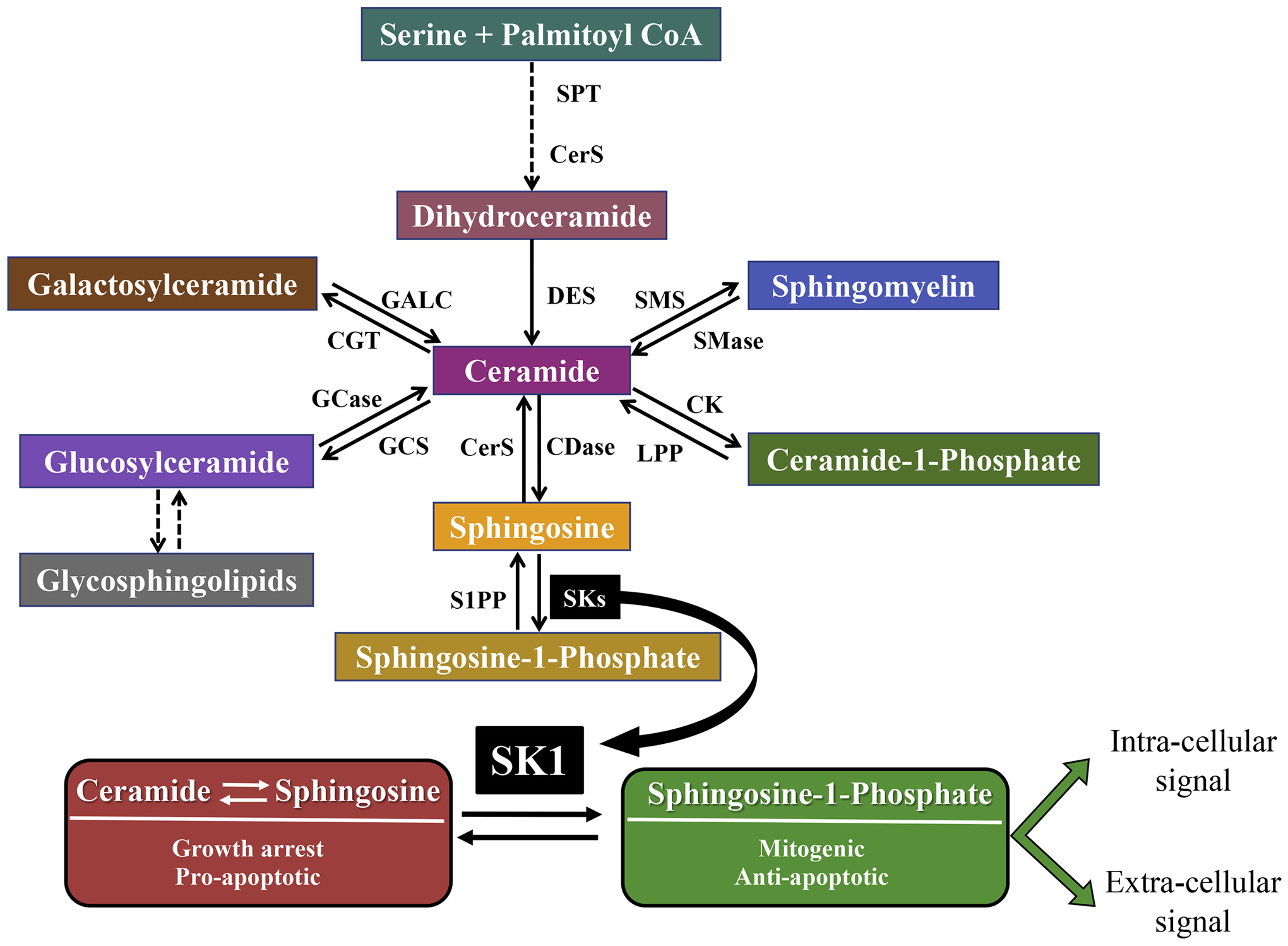
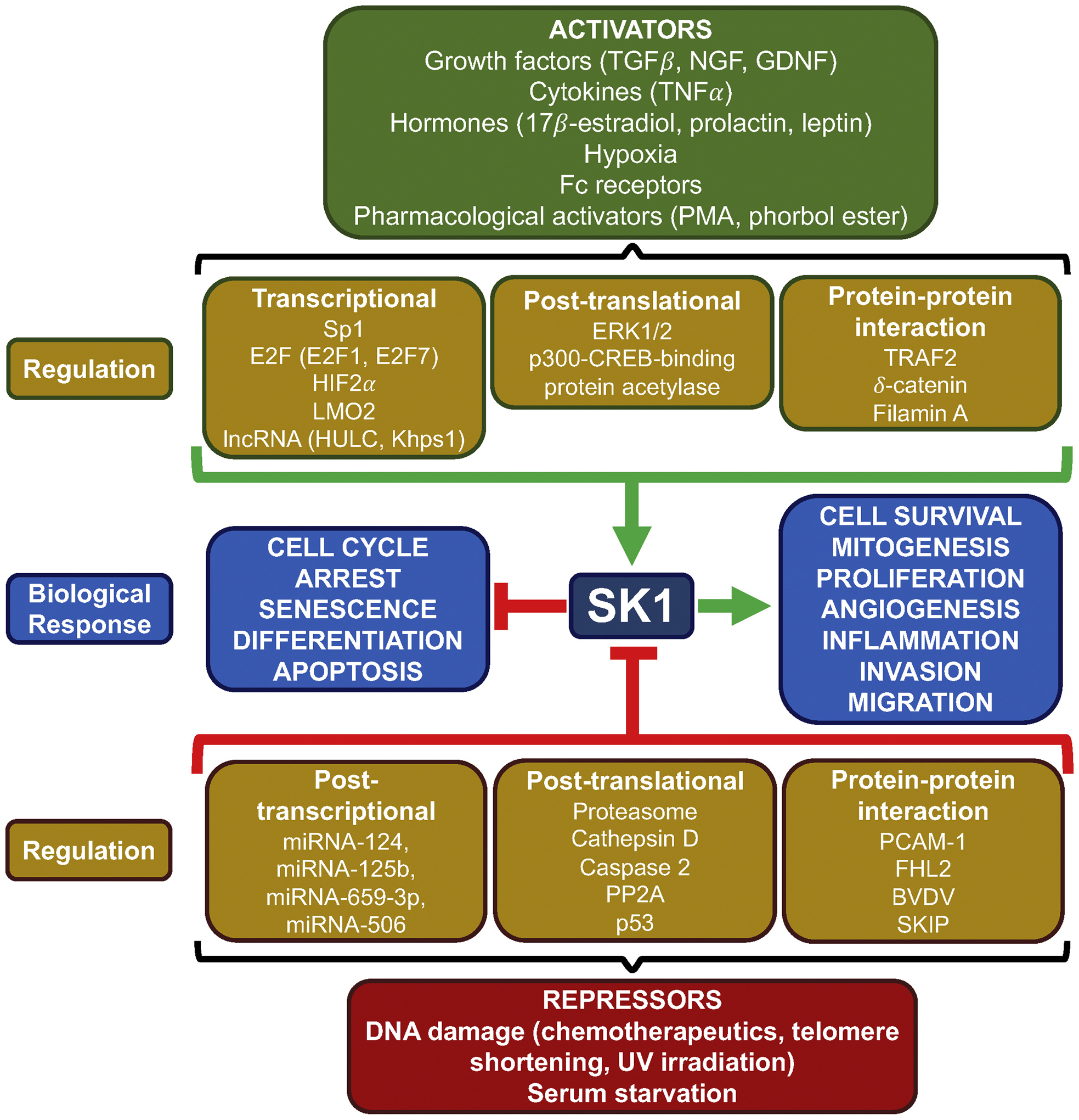
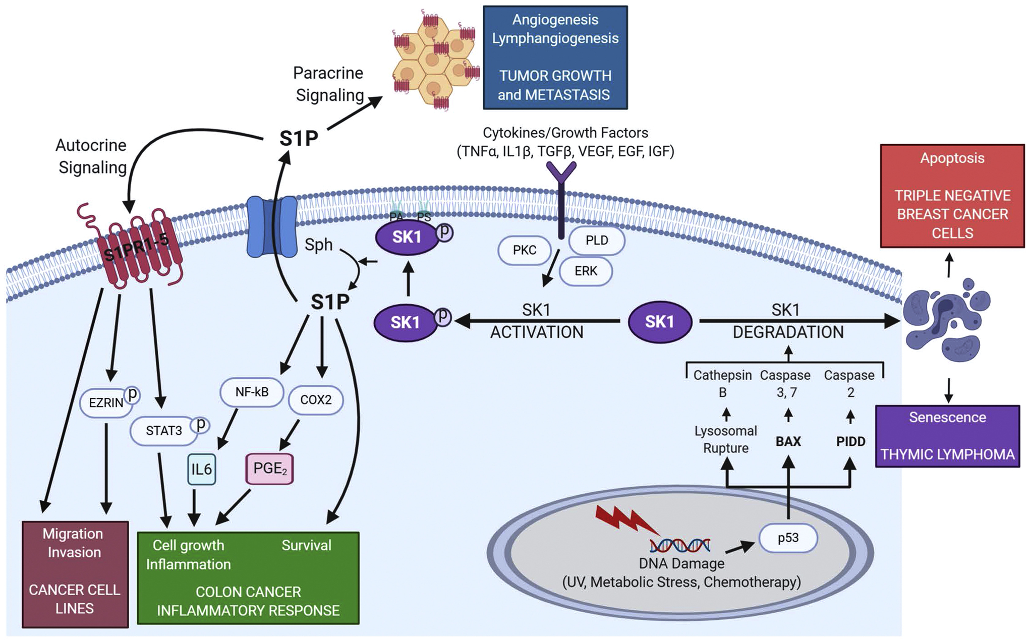
Sphingolipids and their synthetic enzymes have emerged as critical mediators in numerous diseases including inflammation, aging, and cancer. One enzyme in particular, sphingosine kinase (SK) and its product sphingosine-1-phosphate (S1P), has been extensively implicated in these processes. SK catalyzes the phosphorylation of sphingosine to S1P and exists as two isoforms, SK1 and SK2. In this review, we will discuss the contributions from the laboratory of Dr. Lina M. Obeid that have defined the roles for several bioactive sphingolipids in signaling and disease with an emphasis on her work defining SK1 in cellular fates and pathobiologies including proliferation, senescence, apoptosis, and inflammation.


Figures

Fig. 1.
Fig. 1.
SK1: a critical point in sphingolipid metabolism. Sphingolipids are generated de novo via the condensation of serine and palmitoyl-CoA. Ceramide is formed after stepwise reduction, acylation and desaturation processes and can be used to generate many different sphingolipids. Ceramidases are required to generate sphingosine, which can then be phosphorylated by one of two sphingosine kinases (SK1 and SK2) to form S1P. SK1 activity is critical in regulating the balance between two groups of sphingolipids with opposite functions. Ceramide and sphingosine have been shown to cause growth arrest and to activate pro-apoptotic signaling, while S1P has been demonstrated to exert mitogenic and anti-apoptotic properties. S1P can act at the intracellular level, through the binding with different proteins, or upon export, can function as a ligand for one of 5 S1PRs triggering intracellular signaling cascades. SK1 participates in several processes such as cell survival, angiogenesis, lymphocyte trafficking, migration, inflammation and chemotherapeutic resistance, through its product S1P (or the alteration of ceramide and/or sphingosine levels). SPT, serine palmitoyltransferase; CerS, ceramide synthase; DES, desaturase; CK, ceramide kinase; LPP, lipid phosphate phosphatase, CDase, ceramidase, SMS, sphingomyelin synthase; SMase, sphingomyelinase; CGT, ceramide galactosyl transferase; GALC, galactosyl ceramidase; GCS, glucosylceramide synthase; GCase, glucocerebrosidase; S1PP, S1P phosphatase.
Fig. 2.
Fig. 2.
Regulation of SK1. SK1 is positively regulated by a wide range of signaling molecules including growth factors, cytokines and hormones as well as by pharmacological compounds, Fc receptors and hypoxia. These activators drive SK1 activity through various signaling pathways resulting in transcriptional upregulation, post-translational modifications including phosphorylation, or by protein-protein interactions. Increased SK1 activity is primarily associated with cell survival, mitogenesis, proliferation, angiogenesis, inflammation, invasion, or migration in a context-dependent manner. Alternatively, SK1 is negatively regulated by DNA damage induced by chemotherapeutics, UV irradiation, telomere shortening as well as serum starvation. Suppression of SK1 activity can be mediated through post-transcriptional repression by miRNA, post-translational proteasomal degradation, proteolysis via other proteases, dephosphorylation, or through interaction with proteins. Loss of SK1 activity is primarily associated with cell cycle arrest, senescence, apoptosis, and differentiation. PMA, phorbol 12-myristate 13-acetate; ERK, extracellular regulated kinase; TRAF2, TNF receptor-associated factor 2; PP2A, Protein phosphatase 2A; PECAM-1, platelet endothelial cell adhesion molecule 1; FHL2, four-and-a-half LIM domain 2; BVDV, bovine viral diarrhea virus.
Fig. 3.
Fig. 3.
SK1 signaling and associated biological responses. SK1 is activated by a myriad of extra cellular signals, such as growth factors and cytokines. This in turn generates S1P which can be transported out of the cell to act in an autocrine manner where it can modulate many signaling pathways and responses, including increased migration and invasion of cancer cells through the phosphorylation of Ezrin and ERM (ezrin, radixin, moesin) proteins. S1P can also act in paracrine manner driving angiogenesis and lymphangiogenesis leading to tumor growth and metastasis. SK1 and S1P also activate inflammatory signaling via NF-kB, STAT3 and COX2, signals that are critical for colitis and colon cancer. Intracellular signaling via effectors such as PKC, PLD and ERK can result in the phosphorylation and membrane association of SK1, resulting in increased cell survival and proliferation. PLD also produces phosphatidic acid (PA) which helps anchor SK1 in the membrane. DNA damaging agents can also lead to the degradation of SK1 via proteases leading to apoptosis and senescent in cancer cells. *Figure created with BioRender.com.
Fig. 4.
Fig. 4.
In silico surface binding analysis of SK1. A and B: Electrostatic potential maps were generated using Chimera software using the Adaptive Poisson-Boltzman Solver. The scale represents kcal·mol−1 where blue represents positively charged areas and red represents negatively charged areas. C and D: The Kyte-Doolittle scale of hydrophobicity was used to predict hydrophobicity and was mapped to the surface of SK1 where cyan represents hydrophilic areas and magenta represents hydrophobic areas. E and F: Cartoon representation of SK1 with residues of the hydrophobic patch and electrostatic patch represented as magenta and blue sticks, respectively. Protein Data Bank identification number 3VZB [32] used for all analyses. This figure was originally published in the Journal of Lipid Research. Pulkoski-Gross M. J., Jenkins, M.L., Truman, J.P., Salama, M.F., Clarke, C. J., Burke, J.E., Hannun, Y.A., Obeid L.M. An intrinsic lipid-binding interface controls sphingosine kinase 1 function. J Lipid Res. 2018; 59:462–474. © the American Society for Biochemistry and Molecular Biology.

References

    1. Hannun YA, Obeid LM, Principles of bioactive lipid signalling: lessons from sphingolipids, Nat. Rev. Mol. Cell Biol 9 (2008) 139–150. - PubMed
    1. Zitomer NC, Mitchell T, Voss KA, Bondy GS, Pruett ST, Gamier-Amblard EC, Liebeskind LS, Park H, Wang E, Sullards MC, Merrill AH Jr., Riley RT, Ceramide synthase inhibition by fumonisin B1 causes accumulation of 1-deoxysphinganine: a novel category of bioactive 1-deoxysphingoid bases and 1-deoxydihydroceramides biosynthesized by mammalian cell lines and animals, J. Biol. Chem 284 (2009) 4786–4795. - PMC - PubMed
    1. Hannun YA, Obeid LM, Many ceramides J. Biol. Chem 286 (2011) 27855–27862. - PMC - PubMed
    1. Salama MF, Carroll B, Adada M, Pulkoski-Gross M, Hannun YA, Obeid LM, A novel role of sphingosine kinase-1 in the invasion and angiogenesis of VHL mutant clear cell renal cell carcinoma, FASEB J 29 (2015) 2803–2813. - PMC - PubMed
    1. Anelli V, Gault CR, Cheng AB, Obeid LM, Sphingosine Kinase 1 Is Upregulated during Hypoxia in U87MG glioma cells: role of Hypoxia-inducible factors 1 and 2, J. Biol. Chem 283 (2008) 3365–3375. - PubMed

Publication types

MeSH terms