. 2021 Jan;14(1):100975.
doi: 10.1016/j.tranon.2020.100975. Epub 2020 Dec 5.

Fatty acid binding protein-4 promotes alcohol-dependent hepatosteatosis and hepatocellular carcinoma progression

Affiliations

Fatty acid binding protein-4 promotes alcohol-dependent hepatosteatosis and hepatocellular carcinoma progression

Neha Attal et al. Transl Oncol. 2021 Jan.

Abstract

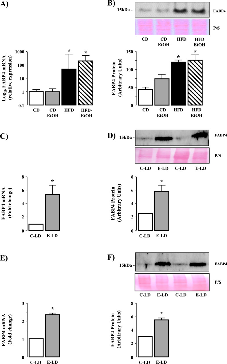
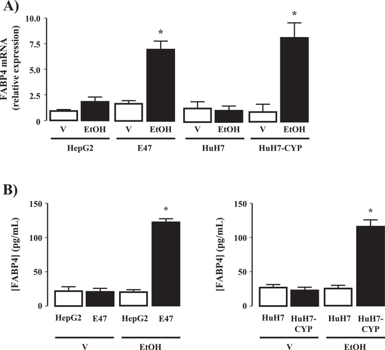
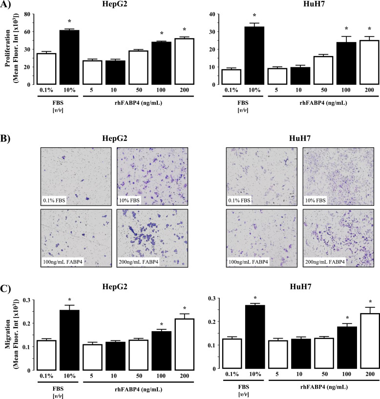
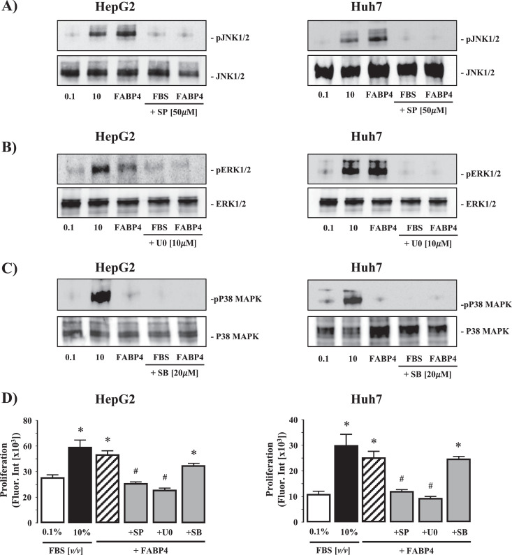
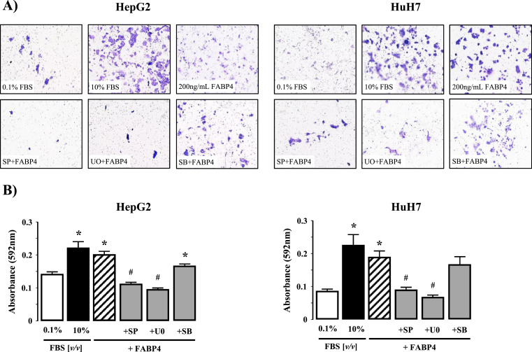
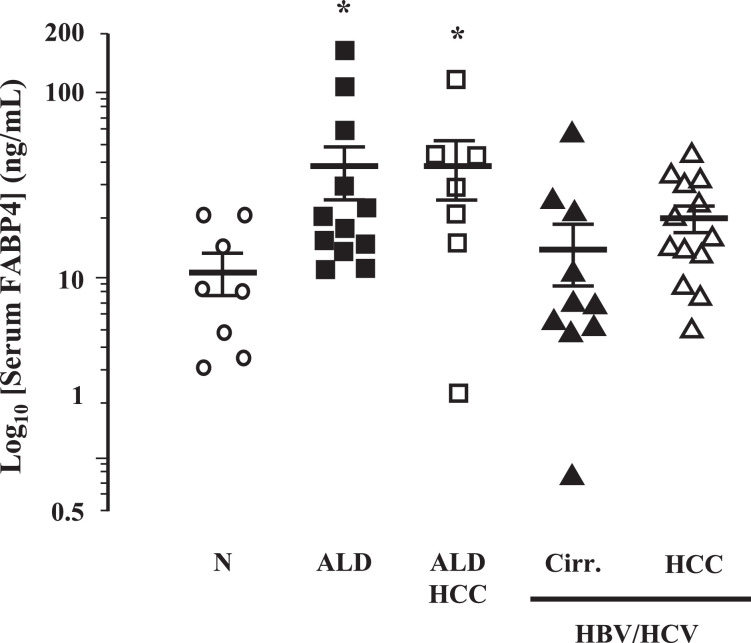
Fatty liver disease (hepatosteatosis) is a common early pathology in alcohol-dependent and obese patients. Fatty acid binding protein-4 (FABP4) is normally expressed in adipocytes and macrophages and functions as a regulator of intracellular lipid movement/storage. This study sought to investigate hepatic FABP4 expression and function in alcoholic liver disease (ALD) and hepatocellular carcinoma (HCC). Using chronic ethanol fed mouse models and patient samples FABP4 expression was analyzed. Human HCC cells, and HCC cells transfected to express CYP2E1, were exposed to ethanol and analyzed for FABP4 expression, or exposed to rhFABP4 (in the absence/presence of ERK, p38-MAPK or JNK1/2 inhibitors) and cell proliferation and migration measured. Hepatosteatotic-ALD mouse models exhibited increased hepatic FABP4 mRNA and protein levels, with FABP4 expression confirmed in hepatocytes. In HCC cells, CYP2E1-dependent ethanol metabolism induced FABP4 expression in vitro and exogenous rhFABP4 stimulated proliferation and migration, effects abrogated by ERK and JNK1/2 inhibition. Increased FABP4 was also detected in ALD/ALD-HCC patients, but not patients with viral hepatitis/HCC. Collectively these data demonstrate ethanol metabolism induces hepatic FABP4 expression and FABP4 promotes hepatoma cell proliferation/migration. These data suggest liver-derived FABP4 may be an important paracrine-endocrine factor during hepatic foci expansion and/or hepatoma progression in the underlying setting of ALD.

Keywords: Alcohol; Fatty acid binding protein 4; Hepatocellular carcinoma; Hepatosteatosis; Liver.


Conflict of interest statement

Declaration of Competing Interest None of the authors have any conflicts of interest to declare.

Figures

Fig 1
Fig. 1
High fat diet and chronic ethanol feeding dysregulate hepatic gene expression. Excised livers were processed for total RNA and gene array analyses performed. (A) Examination of a heat map demonstrating differentially expressed genes (top 200 in magnitude) between mice fed control diet with drinking water (CD) or ethanol in drinking water (CD-EtOH) and animals on high fat diet drinking water (HFD) or HFD and ethanol in drinking water (HFD-EtOH). (B) Venn Diagram identifying genes dysregulated between CD versus HFD and CD versus HFD-EtOH.
Fig 2
Fig. 2
FABP4 is upregulated in models of obesity and ethanol consumption and expressed in hepatocytes from ethanol fed mice. (A) FABP4 mRNA expression in liver tissue isolated from mice fed control diet with drinking water (CD) or ethanol in drinking water (CD-EtOH), and mice on high fat diet drinking water (HFD) or HFD and ethanol in drinking water (HFD-EtOH). *P<0.05 versus CD and CD-EtOH. (B) Western blot analysis of FABP4 protein expression in liver tissue isolated from CD, CD-EtOH, HFD, HFD-EtOH (upper panel) and cumulative densitometric analysis of FABP4 protein expression (lower panel). *P<0.05 versus CD and CD-EtOH. (C) FABP4 mRNA expression in liver tissue from mice maintained on control Lieber-DeCarli liquid diet (C-LD) or ethanol containing Lieber-DeCarli liquid diet (E-LD). *P<0.05 versus C-LD. (D) Western blot analysis of FABP4 protein expression in liver tissue from mice maintained on C-LD or E-LD (upper panel) and cumulative densitometric analysis of FABP4 protein expression (lower panel). *P<0.05 versus C-LD. (E) FABP4 mRNA expression in isolated hepatocytes from mice maintained on C-LD or E-LD. *P<0.05 versus C-LD. (F) Western blot analysis of FABP4 protein expression in isolated hepatocytes from mice maintained on C-LD or E-LD (upper panel) and cumulative densitometric analysis of FABP4 protein expression (lower panel). *P<0.05 versus C-LD.
Fig 3
Fig. 3
Cytochrome P4502E1 (CYP2E1) dependent ethanol metabolism induces FABP4 expression in vitro. (A) FABP4 mRNA expression in HepG2 and HuH7 cells and HepG2 and HuH7 stably transfected to express CYP2E1(E47 and HuH7-CYP) following exposure to vehicle (V; 0.1mL phosphate buffered saline) or 100 mM ethanol (EtOH; 48h). *P<0.05 EtOH versus V, transfected versus non-transfected, N = 4 independent experiments. FABP4 secretion into culture medium from (B) HepG2/E47 and (C) HuH7/HuH7-CYP cells following exposure to V or 50 mM EtOH (48h). *P<0.05 EtOH versus V transfected versus non-transfected, N = 4 independent experiments.
Fig. 4
Fig. 4
rhFABP4 stimulates hepatoma cell proliferation and migration in vitro. (A) HepG2 and HuH7 cells were exposed to rhFABP4 (0–200 ng/mL, 48h) and assayed for cell proliferation. Negative and positive controls were 0.1% [v/v] and 10% [v/v] fetal bovine serum (FBS). *P<0.05 versus 0.1% [v/v] FBS, N = 4 independent experiments. (B) Representative images of HepG2 and HuH7 cell migration following 48h in the presence of 0.1% [v/v] FBS (control), 10% [v/v] FBS, 100 ng/mL or 200 ng/mL rhFABP4. (C) Cumulative analysis of HepG2 and HuH7 cell migration (crystal violet mean fluorescent intensity) following 48h in the presence of 0.1% [v/v] FBS (control), 10% [v/v] FBS, or rhFABP4 (5–200 ng/mL). *P<0.05 versus 0.1% [v/v] FBS and 5 ng/mL rhFABP4, N = 4 independent experiments.
Fig 5
Fig. 5
Intracellular signaling pathways involved in rhFABP4-stimulated hepatoma cell proliferation and migration in vitro. (A) Representative Western blot analysis of phosphorylated/total JNK1/2 (pJNK1/2 - JNK1/2) in HepG2 and HuH7 HCC cells following exposure to 0.1 or 10% (v/v) fetal bovine serum (FBS), rhFABP4 (200 ng/mL), or 10% (v/v) FBS/rhFABP4 in the presence of SP 600,125 [SP; JNK inhibitor] (50 μM). (B) Phosphorylated/total ERK1/2 (pERK 1/2 - ERK 1/2) in HepG2 and HuH7 HCC cells following exposure to FBS, rhFABP4 (200 ng/mL), or FBS/rhFABP4 in the presence of U0126 [U0; ERK1/2 inhibitor] (10 μM). (C) Phosphorylated/total p38 mitogen-activate protein kinase (pp38MAPK - p38MAPK) in HepG2 and HuH7 HCC cells following exposure to FBS, rhFABP4 (100 ng/mL), or FBS/rhFABP4 in the presence of SB 203,589 [SB; p38MAPK inhibitor] (20 μM). D) HepG2 and HuH7 cells were pretreated with SB, U0, or SP for 60 min prior to exposure to exogenous rhFABP4 (200 ng/mL; 24 h) and assayed for proliferation. Negative and positive controls were 0.1% [v/v] and 10% [v/v] FBS respectively. *P<0.05 versus 0.1% [v/v] FBS, #P<0.05 versus rhFABP4, N = 4.
Fig 6
Fig. 6
(A) Representative images of HepG2 and HuH7 cell migration following exposure to 0.1 or 10% (v/v) fetal bovine serum (FBS), rhFABP4 (200 ng/mL), or 10% (v/v) FBS/rhFABP4 in the presence of SP 600,125 [SP; JNK inhibitor] (50 μM). U0126 [U0; ERK1/2 inhibitor] (10 μM), or SB 203,589 [SB; p38MAPK inhibitor] (20 μM). (B) Cumulative analysis of HepG2 and HuH7 cell migration (mean crystal violet fluorescent intensity). *P<0.05 versus 0.1% [v/v] FBS, #P<0.05 versus rhFABP4, N = 4.
Fig. 7
Fig. 7
Serum FABP4 is elevated in ALD patients. Serum isolated from whole blood was collected from human subjects with normal livers (Norm, N = 8), patients with alcoholic liver disease (ALD; N = 12) or HCC caused by ALD (ALD-HCC; N = 7), or patients with cirrhosis caused by viral hepatitis (HBV/HCV-Cirr; N = 10) and viral hepatitis associated HCC (HBV/HCV-HCC; N = 13) and assayed for FABP4 levels. *P< 0.05 versus Norm.

References

    1. Yang J.D., Hainaut P., Gores G.J., Amadou A., Plymoth A., Roberts L.R. A global view of hepatocellular carcinoma: trends, risk, prevention and management. Nat. Rev. Gastroenterol. Hepatol. 2019;16:589–604. - PMC - PubMed
    1. Kumar R., Priyadarshi R.N., Anand U. Non-alcoholic fatty liver disease: growing burden, adverse outcomes and associations. J. Clin. Transl. Hepatol. 2020;8:76–86. - PMC - PubMed
    1. McCullough A.K., Lloyd R.S. Mechanisms underlying aflatoxin-associated mutagenesis - Implications in carcinogenesis. DNA Repair (Amst.) 2019;77:76–86. - PMC - PubMed
    1. McKillop I.H., Girardi C.A., Thompson K.J. Role of fatty acid binding proteins (FABPs) in cancer development and progression. Cell Signal. 2019;62 - PubMed
    1. Sagnelli E., Macera M., Russo A., Coppola N., Sagnelli C. Epidemiological and etiological variations in hepatocellular carcinoma. Infection. 2020;48:7–17. - PubMed