Review
. 2020 Dec 6;9(12):2620.
doi: 10.3390/cells9122620.

HIGD-Driven Regulation of Cytochrome c Oxidase Biogenesis and Function

Affiliations
Review

HIGD-Driven Regulation of Cytochrome c Oxidase Biogenesis and Function

Alba Timón-Gómez et al. Cells. .

Abstract

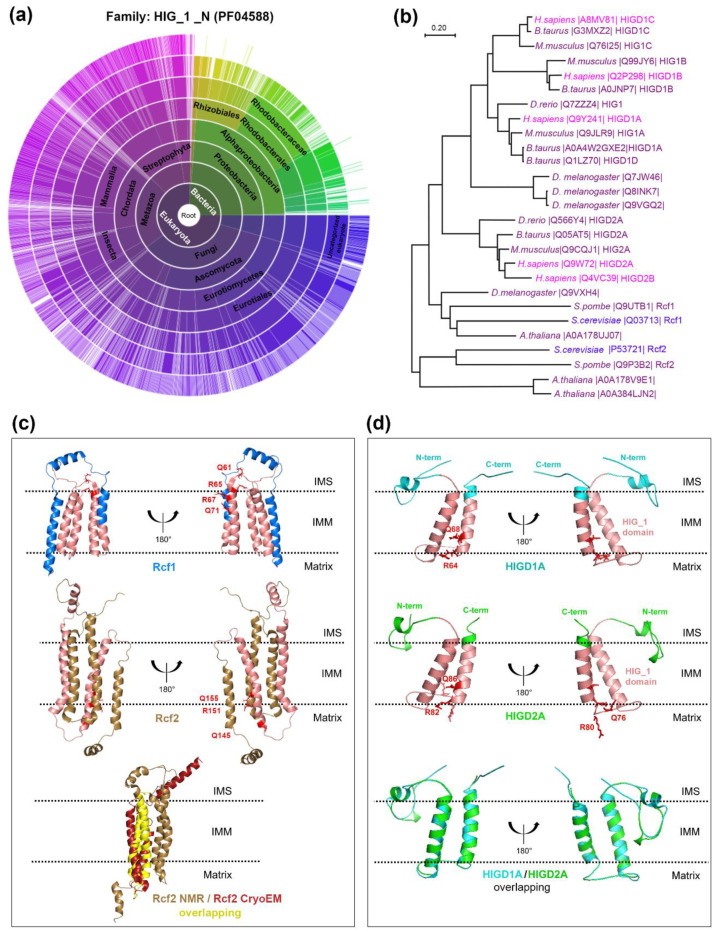
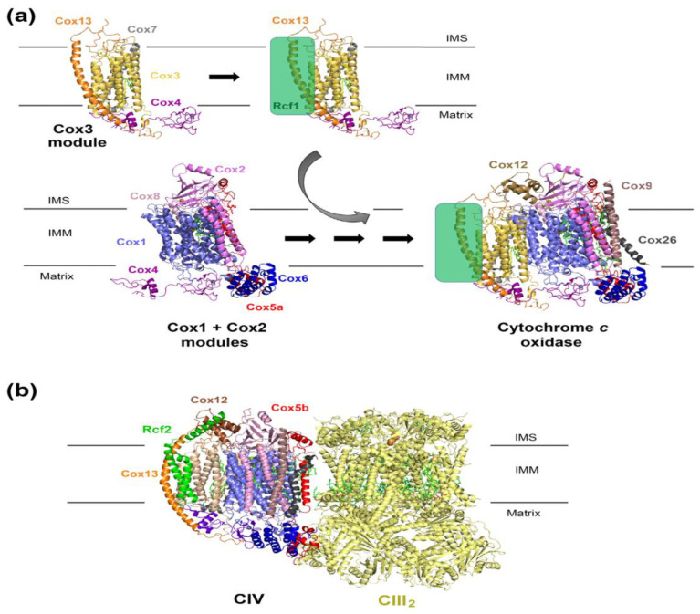
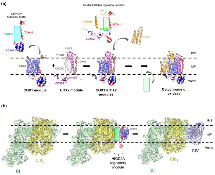
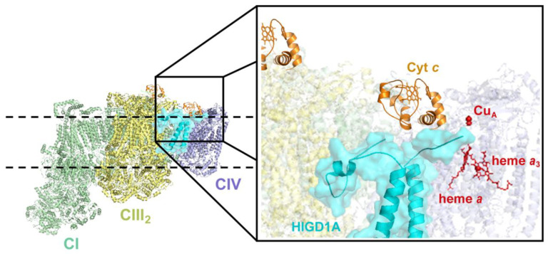
The biogenesis and function of eukaryotic cytochrome c oxidase or mitochondrial respiratory chain complex IV (CIV) undergo several levels of regulation to adapt to changing environmental conditions. Adaptation to hypoxia and oxidative stress involves CIV subunit isoform switch, changes in phosphorylation status, and modulation of CIV assembly and enzymatic activity by interacting factors. The latter include the Hypoxia Inducible Gene Domain (HIGD) family yeast respiratory supercomplex factors 1 and 2 (Rcf1 and Rcf2) and two mammalian homologs of Rcf1, the proteins HIGD1A and HIGD2A. Whereas Rcf1 and Rcf2 are expressed constitutively, expression of HIGD1A and HIGD2A is induced under stress conditions, such as hypoxia and/or low glucose levels. In both systems, the HIGD proteins localize in the mitochondrial inner membrane and play a role in the biogenesis of CIV as a free unit or as part as respiratory supercomplexes. Notably, they remain bound to assembled CIV and, by modulating its activity, regulate cellular respiration. Here, we will describe the current knowledge regarding the specific and overlapping roles of the several HIGD proteins in physiological and stress conditions.

Keywords: HIGD1A; HIGD2A; Hypoxia Inducible Gene Domain; Rcf1; Rcf2; cytochrome c oxidase; mitochondrial respiratory chain complex IV.


Conflict of interest statement

The authors declare no conflict of interest.

Figures

Figure 1
Figure 1
Distribution of HIG_1_N (PF04588) domain proteins across species. (a) Modified “sunburst” visualization of the taxonomic lineage distribution of 1911 different species that in total have 3472 protein sequences containing the hypoxia-inducible gene (HIG)-1-N domain (Pfam ID: PF04588). The graph shows each node in the tree as a separate arc, arranged radially with the superkingdoms at the center and the species arrayed around the outermost ring. The graph was generated with tools from pfam.xfam.org hosted by the European Bioinformatics Institute at the European Molecular Biology Laboratory (EMBL-EBI). Yellow-green colors represent different types of bacteria, and purple color represent eukaryotes. (b) Phylogenetic tree of Hypoxia-inducible gene domain (HIGD) proteins across eukaryotic species. The evolutionary history was inferred using the Neighbor-Joining method. We show the optimal tree with the sum of branch length = 8.97673253. The tree was drawn to scale, with branch lengths in the same units as those of the evolutionary distances used to infer the phylogenetic tree. The evolutionary distances were computed using the Poisson correction method and are in the units of the number of amino acid substitutions per site. This analysis involved 27 amino acid sequences obtained from UniProt and entered manually. All ambiguous positions were removed for each sequence pair (pairwise deletion option). There were a total of 253 positions in the final dataset. Evolutionary analyses were conducted in MEGA X [42,43]. (c) Solution NMR structures of S. cerevisiae respiratory supercomplex factors Rcf1 and Rcf2. The top panel shows ribbon diagrams of the Rcf1 structure (Protein database -PDB- code 5NF8), with the HIG_1 domain in salmon. The middle panel shows the Rcf2 structure (PDB 6LUL) [44], with the HIG_1 domain in salmon. The QRRQ motifs in Rcf1 and Rcf2 are marked in red. The lower panel shows the overlapping of the NMR structure of Rcf2 (PDB 6LUL) (in gold) and the partial cryo-EM structure for Rcf2 as bound to hypoxic respiratory supercomplex (SC) CIII2+CIV (PDB 6T15) [34] (in red). The Rcf2 transmembrane helices resolved by NMR that overlap with the Rcf2 cryo-electron microscopy (cryo-EM) structure are presented in yellow. A prominent difference between the two structures relates to the C-terminal α-helix, shown to protrude into the intermembrane space by cryo-EM. (d) Solution NMR structures of human HIGD1A and HIGD2A. The top panel shows ribbon diagrams of the structure of HIGD1A (PDB 2LOM), with the HIG_1 domain in salmon. HIGD1A is a type 1 HIGD protein, with a truncated QRRQ motif, labeled in red. The middle panel presents ribbon diagrams of the predicted structure of HIGD2A. The structure was predicted in silico using Swiss-Model based on the alignment with HIGD1A, with 0.75 coverage of the sequence [45,46,47]. Residues of the QRRQ motif are labeled in red and HIG_1 domain, in salmon. The bottom panel depicts a superposition view of HIGD1A and HIGD2A structures generated by PyMOL. IMS: intermembrane space. IMM: inner mitochondrial membrane.
Figure 2
Figure 2
Proposed role of Rcf1 in the regulation of S. cerevisiae cytochrome c oxidase assembly and activity. (a) Schematic view of the yeast S. cerevisiae modular cytochrome c oxidase assembly pathway [21], depicting the role of Rcf1. Rcf1 interacts with the Cox3 and Cox13 in the Cox3-assembly module and promotes its incorporation into the CIV assembly line to join the assembled Cox1 + Cox2 module. The presence of Rcf1 facilitates the incorporation of Cox12. The figure was generated with PyMOL using the S. cerevisiae cytochrome c oxidase cryo-EM structure PDB 6YMY [57]. Rcf1 was incorporated as a green box, interacting with Cox3 and Cox13 as shown by biochemical studies. (b) Structure of the S. cerevisiae hypoxic respiratory SC III2–IV containing the Cox5 hypoxic isoform Cox5b and Rcf2 (PDB 6T15) [34].
Figure 3
Figure 3
Roles of HIGD1A and HIGD2A in cytochrome c oxidase assembly. (a) Schematic view of the mammalian modular cytochrome c oxidase assembly pathway. HIGD1A is present in an early assembly cluster, together with the COX1 module proteins COX4-1 and COX5A. HIGD2A forms a 50 kDa regulatory module with COX4-1, COX5B, and COX6A1, which binds to newly synthesized COX3 to promote its binding to the previously assembled COX1 + COX2 module. HIGD1A and HIGD2A are represented by a blue and a green box, respectively, due to the lack of structural information. (b) The HIGD2A regulatory module plays a role in supercomplexed cytochrome c oxidase assembly, by interacting with SC I+III2 and facilitating the incorporation of the remaining CIV subunits. The figure was generated with PyMOL and Adobe Photoshop using the structure of the CI-CIII2-CIV respirasome from ovine heart mitochondria [81] (PDB 5J4Z). IMS: intermembrane space. IMM: inner mitochondrial membrane. CI: complex I. CIII2: complex III dimer. CIV: complex IV.
Figure 4
Figure 4
HIGD1A interacts with CIV and enhances its activity. Following a proposed model [31,70], overexpressed HIGD1A interacts with cytochrome c and CIV and produces a structural change in the CIV heme a active center, acting as a positive modulator of the enzyme. The figure was generated with PyMOL and Adobe Photoshop using the structure of the CI-CIII2-CIV respirasome from ovine (Ovis aries) heart mitochondria [81](PDB 5J4Z). IMS, intermembrane space. IMM, inner mitochondrial membrane. CI, complex I. CIII2, complex III dimer. CIV, complex IV.

References

    1. Kadenbach B. Complex IV—The regulatory center of mitochondrial oxidative phosphorylation. Mitochondrion. 2020 in press. - PubMed
    1. Timon-Gomez A., Nyvltova E., Abriata L.A., Vila A.J., Hosler J., Barrientos A. Mitochondrial cytochrome c oxidase biogenesis: Recent developments. Semin. Cell Dev. Biol. 2018;76:163–178. doi: 10.1016/j.semcdb.2017.08.055. - DOI - PMC - PubMed
    1. Pierron D., Wildman D.E., Huttemann M., Markondapatnaikuni G.C., Aras S., Grossman L.I. Cytochrome c oxidase: Evolution of control via nuclear subunit addition. Biochim. Biophys. Acta Bioenerg. 2012;1817:590–597. doi: 10.1016/j.bbabio.2011.07.007. - DOI - PMC - PubMed
    1. Huttemann M., Kadenbach B., Grossman L.I. Mammalian subunit IV isoforms of cytochrome c oxidase. Gene. 2001;267:111–123. doi: 10.1016/S0378-1119(01)00385-7. - DOI - PubMed
    1. Poyton R.O., Burke P.V. Oxygen regulated transcription of cytochrome c and cytochrome c oxidase genes in yeast. Biochim. Biophys. Acta Bioenerg. 1992;1101:252–256. doi: 10.1016/S0005-2728(05)80033-7. - DOI - PubMed

Publication types

MeSH terms

LinkOut - more resources