

Influence of PDA Coating on the Structural, Optical and Surface Properties of ZnO Nanostructures
- PMID: 33291264
- PMCID: PMC7762110
- DOI: 10.3390/nano10122438
Influence of PDA Coating on the Structural, Optical and Surface Properties of ZnO Nanostructures
Abstract
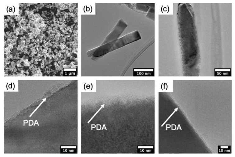
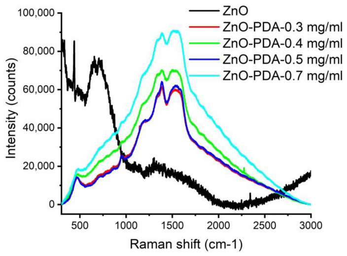
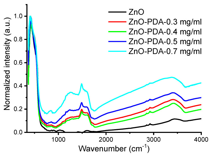
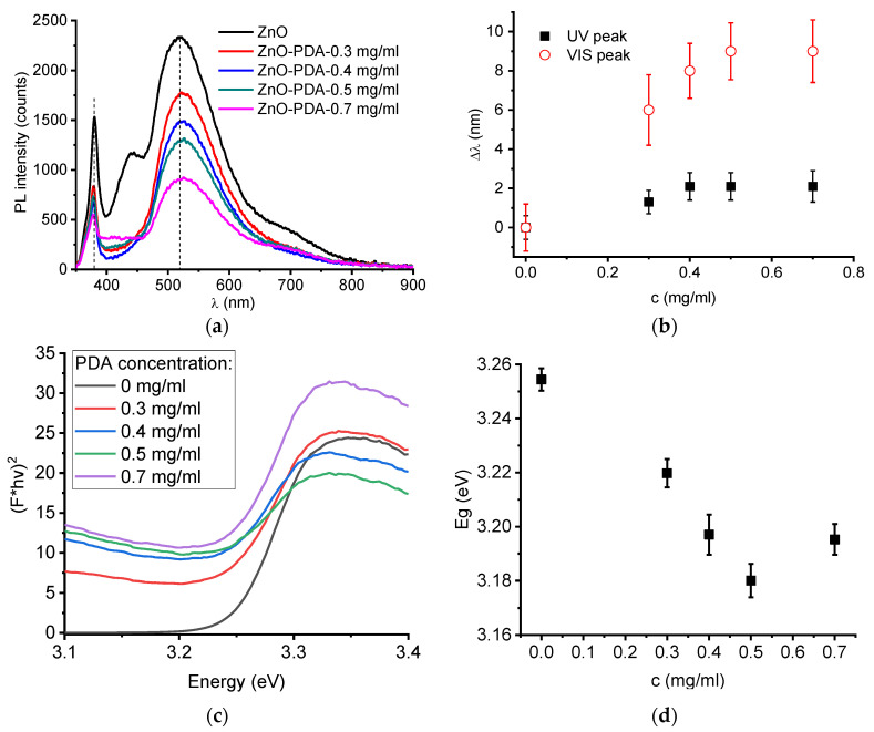
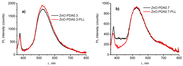
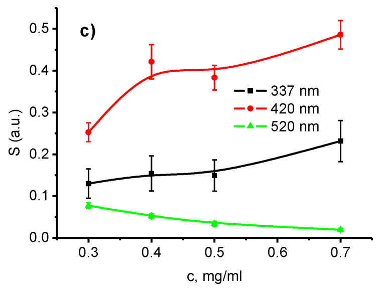
Polydopamine (PDA) is a new biocompatible material, which has prospects in biomedical and sensor applications. Due to functional groups, it can host wide range of biomolecules. ZnO nanostructures are well known templates for optical sensors and biosensors. The combination of ZnO and PDA results in a change of optical properties of ZnO-PDA composites as a shift of photoluminescence (PL) peaks and PL quenching. However, to date, the effect of the PDA layer on fundamental properties of ZnO-PDA nanostructures has not been studied. The presented paper reports on optical and surface properties of novel ZnO-PDA nanocomposites. PDA layers were chemically synthesized on ZnO nanostructures from different solution concentrations of 0.3, 0.4, 0.5 and 0.7 mg/mL. Structure, electronic and optical properties were studied by SEM, Raman, FTIR, diffuse reflectance and photoluminescence methods. The Z-potential of the samples was evaluated in neutral pH (pH = 7.2). The response of the samples towards poly-l-lysine adsorption, as a model molecule, was studied by PL spectroscopy to evaluate the correlation between optical and surface properties. The role of the PDA concentration on fundamental properties was discussed.
Keywords: ZnO–polydopamine nanocomposites; fundamental properties; optical sensors.
Conflict of interest statement
The authors declare no conflict of interest.
Figures
Similar articles
-
Application of Polydopamine Functionalized Zinc Oxide for Glucose Biosensor Design.Polymers (Basel). 2021 Aug 30;13(17):2918. doi: 10.3390/polym13172918. Polymers (Basel). 2021. PMID: 34502958 Free PMC article.
-
Synthesis and photoluminescence properties of hybrid 1D core-shell structured nanocomposites based on ZnO/polydopamine.RSC Adv. 2020 Aug 12;10(50):29751-29758. doi: 10.1039/d0ra04829a. eCollection 2020 Aug 10. RSC Adv. 2020. PMID: 35518237 Free PMC article.
-
Porous Silicon-Zinc Oxide Nanocomposites Prepared by Atomic Layer Deposition for Biophotonic Applications.Materials (Basel). 2020 Apr 24;13(8):1987. doi: 10.3390/ma13081987. Materials (Basel). 2020. PMID: 32344562 Free PMC article.
-
ZnO-based nanostructured electrodes for electrochemical sensors and biosensors in biomedical applications.Biosens Bioelectron. 2019 Sep 15;141:111417. doi: 10.1016/j.bios.2019.111417. Epub 2019 Jun 8. Biosens Bioelectron. 2019. PMID: 31202187 Review.
-
Polydopamine-based plasmonic nanocomposites: rational designs and applications.Chem Commun (Camb). 2024 Mar 12;60(22):2982-2993. doi: 10.1039/d3cc05883b. Chem Commun (Camb). 2024. PMID: 38384206 Review.
Cited by
-
Application of Polydopamine Functionalized Zinc Oxide for Glucose Biosensor Design.Polymers (Basel). 2021 Aug 30;13(17):2918. doi: 10.3390/polym13172918. Polymers (Basel). 2021. PMID: 34502958 Free PMC article.
-
Fabrication of Gelatin-ZnO Nanofibers for Antibacterial Applications.Materials (Basel). 2020 Dec 29;14(1):103. doi: 10.3390/ma14010103. Materials (Basel). 2020. PMID: 33383718 Free PMC article.
-
Dual-step photo-induced self-assembled hydrogel for endogenous oral mucosal wound healing.Light Sci Appl. 2025 May 15;14(1):186. doi: 10.1038/s41377-025-01837-7. Light Sci Appl. 2025. PMID: 40368885 Free PMC article.
-
Preparation of an innovative series of respiratory nano-filters using polystyrene fibrous films containing KCC-1 dendrimer and ZnO nanostructures for environmental assessment of SO2, NO2 and CO2.RSC Adv. 2024 Mar 5;14(11):7303-7313. doi: 10.1039/d4ra00176a. eCollection 2024 Feb 29. RSC Adv. 2024. PMID: 38444973 Free PMC article.
-
Laser-Induced Graphitization of Polydopamine on Titania Nanotubes.ACS Appl Mater Interfaces. 2023 Nov 15;15(45):52921-52938. doi: 10.1021/acsami.3c11580. Epub 2023 Nov 1. ACS Appl Mater Interfaces. 2023. PMID: 37915241 Free PMC article.
References
-
- Singh A., Mathur A., Pal D., Sengupta A., Singh R., Chattopadhyay S. Near room temperature atomic layer deposition of ZnO thin films on poly (methyl methacrylate) (PMMA) templates: A study of structure, morphology and photoluminescence of ZnO as an effect of template confinement. Vacuum. 2019;161:398–403. doi: 10.1016/j.vacuum.2019.01.006. - DOI
-
- Viter R., Savchuk M., Iatsunskyi I., Pietralik Z., Starodub N., Shpyrka N., Ramanaviciene A., Ramanavicius A. Analytical, thermodynamical and kinetic characteristics of photoluminescence immunosensor for the determination of Ochratoxin A. Biosens. Bioelectron. 2018;99:237–243. doi: 10.1016/j.bios.2017.07.056. - DOI - PubMed
Grants and funding
LinkOut - more resources
Full Text Sources
