

Serum Metabolomic Profiling of Patients with Non-Infectious Uveitis
- PMID: 33291298
- PMCID: PMC7762156
- DOI: 10.3390/jcm9123955
Serum Metabolomic Profiling of Patients with Non-Infectious Uveitis
Abstract
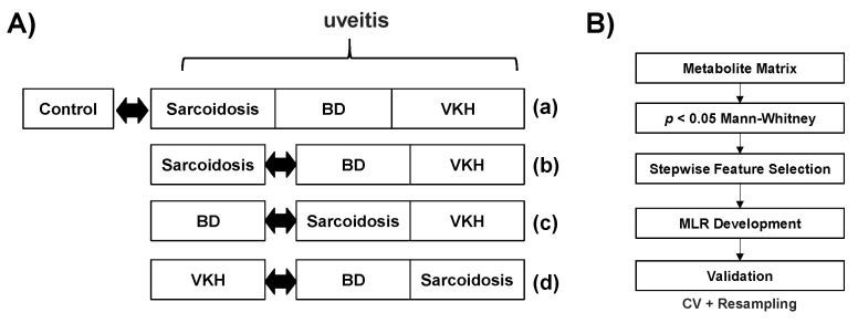
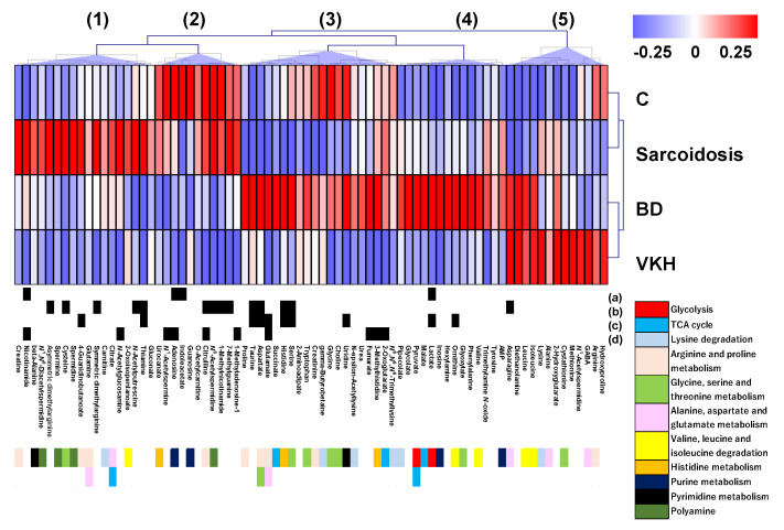
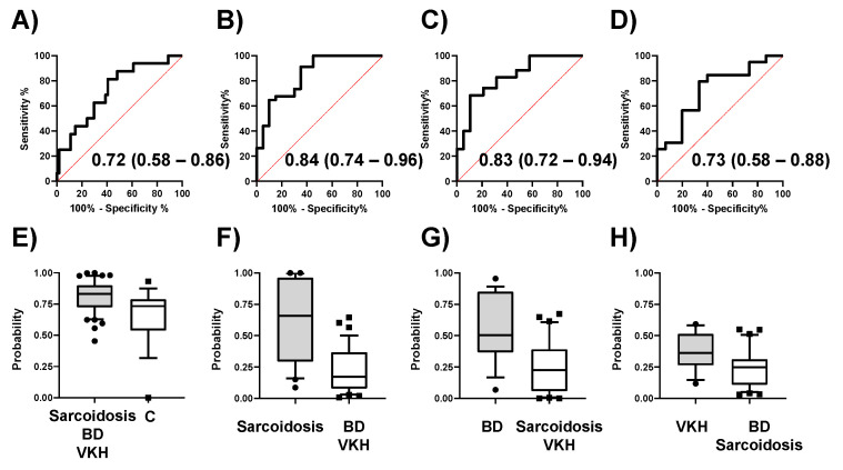
The activities of various metabolic pathways can influence the pathogeneses of autoimmune diseases, and intrinsic metabolites can potentially be used to diagnose diseases. However, the metabolomic analysis of patients with uveitis has not yet been conducted. Here, we profiled the serum metabolomes of patients with three major forms of uveitis (Behҫet's disease (BD), sarcoidosis, and Vogt-Koyanagi-Harada disease (VKH)) to identify potential biomarkers. This study included 19 BD, 20 sarcoidosis, and 15 VKH patients alongside 16 healthy control subjects. The metabolite concentrations in their sera were quantified using liquid chromatography with time-of-flight mass spectrometry. The discriminative abilities of quantified metabolites were evaluated by four comparisons: control vs. three diseases, and each disease vs. the other two diseases (such as sarcoidosis vs. BD + VKH). Among 78 quantified metabolites, 24 kinds of metabolites showed significant differences in these comparisons. Four multiple logistic regression models were developed and validated. The area under the receiver operating characteristic (ROC) curve (AUC) in the model to discriminate disease groups from control was 0.72. The AUC of the other models to discriminate sarcoidosis, BD, and VKH from the other two diseases were 0.84, 0.83, and 0.73, respectively. This study provides potential diagnostic abilities of sarcoidosis, BD, and VKH using routinely available serum samples that can be collected with minimal invasiveness.
Keywords: Behҫet’s disease; Vogt-Koyanagi-Harada disease; biomarker; liquid chromatography-mass spectrometry; metabolomics; sarcoidosis; serum.
Conflict of interest statement
The authors declare that they have no competing interest.
Figures
Similar articles
-
Comprehensive miRNA Analysis Using Serum From Patients With Noninfectious Uveitis.Invest Ophthalmol Vis Sci. 2020 Sep 1;61(11):4. doi: 10.1167/iovs.61.11.4. Invest Ophthalmol Vis Sci. 2020. PMID: 32876691 Free PMC article.
-
Identification of Urine Metabolic Biomarkers for Vogt-Koyanagi-Harada Disease.Front Cell Dev Biol. 2021 Feb 25;9:637489. doi: 10.3389/fcell.2021.637489. eCollection 2021. Front Cell Dev Biol. 2021. PMID: 33718374 Free PMC article.
-
Metabolomic Analysis of Aqueous Humor Identifies Aberrant Amino Acid and Fatty Acid Metabolism in Vogt-Koyanagi-Harada and Behcet's Disease.Front Immunol. 2021 Feb 22;12:587393. doi: 10.3389/fimmu.2021.587393. eCollection 2021. Front Immunol. 2021. PMID: 33732231 Free PMC article.
-
Molecular Genetic Advances in Uveitis.Prog Mol Biol Transl Sci. 2015;134:283-98. doi: 10.1016/bs.pmbts.2015.04.009. Prog Mol Biol Transl Sci. 2015. PMID: 26310161 Review.
-
Catching the therapeutic window of opportunity in early initial-onset Vogt-Koyanagi-Harada uveitis can cure the disease.Int Ophthalmol. 2019 Jun;39(6):1419-1425. doi: 10.1007/s10792-018-0949-4. Epub 2018 Jun 11. Int Ophthalmol. 2019. PMID: 29948499 Review.
Cited by
-
Small molecule metabolites: discovery of biomarkers and therapeutic targets.Signal Transduct Target Ther. 2023 Mar 20;8(1):132. doi: 10.1038/s41392-023-01399-3. Signal Transduct Target Ther. 2023. PMID: 36941259 Free PMC article. Review.
-
Comprehensive Proteomic Profiling of Vitreous Humor in Ocular Sarcoidosis Compared with Other Vitreoretinal Diseases.J Clin Med. 2022 Jun 22;11(13):3606. doi: 10.3390/jcm11133606. J Clin Med. 2022. PMID: 35806888 Free PMC article.
-
Metabolomic profiling of cancer-related fatigue involved in cachexia and chemotherapy.Sci Rep. 2024 Apr 9;14(1):8329. doi: 10.1038/s41598-024-57747-y. Sci Rep. 2024. PMID: 38594321 Free PMC article.
-
Serum metabolomics in pulmonary sarcoidosis: metabolic signatures across prognoses.BMC Pulm Med. 2025 Aug 2;25(1):373. doi: 10.1186/s12890-025-03863-y. BMC Pulm Med. 2025. PMID: 40753209 Free PMC article.
-
Long-Term Mastication Changed Salivary Metabolomic Profiles.Metabolites. 2022 Jul 18;12(7):660. doi: 10.3390/metabo12070660. Metabolites. 2022. PMID: 35888784 Free PMC article.
References
Grants and funding
- 17ae0101016s0904/Japan Agency for Medical Research and Development
- 16K11330/The Ministry of Education, Culture, Sports, Science and Technology of Japan
- 19K09981/The Ministry of Education, Culture, Sports, Science and Technology of Japan
- 19K09959/The Ministry of Education, Culture, Sports, Science and Technology of Japan
LinkOut - more resources
Full Text Sources
