. 2020 Dec 5;21(23):9293.
doi: 10.3390/ijms21239293.

Characterization of AMBN I and II Isoforms and Study of Their Ca2+-Binding Properties

Affiliations

Characterization of AMBN I and II Isoforms and Study of Their Ca2+-Binding Properties

Veronika Vetyskova et al. Int J Mol Sci. .

Abstract

Ameloblastin (Ambn) as an intrinsically disordered protein (IDP) stands for an important role in the formation of enamel-the hardest biomineralized tissue commonly formed in vertebrates. The human ameloblastin (AMBN) is expressed in two isoforms: full-length isoform I (AMBN ISO I) and isoform II (AMBN ISO II), which is about 15 amino acid residues shorter than AMBN ISO I. The significant feature of AMBN-its oligomerization ability-is enabled due to a specific sequence encoded by exon 5 present at the N-terminal part in both known isoforms. In this study, we characterized AMBN ISO I and AMBN ISO II by biochemical and biophysical methods to determine their common features and differences. We confirmed that both AMBN ISO I and AMBN ISO II form oligomers in in vitro conditions. Due to an important role of AMBN in biomineralization, we further addressed the calcium (Ca2+)-binding properties of AMBN ISO I and ISO II. The binding properties of AMBN to Ca2+ may explain the role of AMBN in biomineralization and more generally in Ca2+ homeostasis processes.

Keywords: ameloblastin; biomineralization; calcium binding; intrinsically disordered protein (IDPs); oligomerization.


Conflict of interest statement

The authors declare no conflict of interest.

Figures

Figure 1
Figure 1
(A) Circular dichroism (CD) spectra of ameloblastin full-length isoform I (AMBN ISO I), ameloblastin full-length isoform II (AMBN ISO II) and AMBN with deleted exon 5 (AMBN del E5). Sample before (black) and after (red) addition of 10 mM CaCl2. (B) Analysis of autocorrelation functions (ACF) from dynamic light scattering (DLS) spectra of AMBN ISO I, AMBN ISO II, AMBN del E5, and bovine serum albumin (BSA) as a standard for globular protein. ACF of proteins in the absence or presence of 10mM CaCl2. (C) First derivative of thermal unfolding curves of AMBN ISO I, AMBN ISO II, and AMBN del E5 in the presence or absence of 10 mM CaCl2.
Figure 2
Figure 2
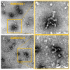
(A) Transmission electron micrographs of AMBN ISO I. Samples were examined at a magnification of 200,000×; scale bar = 100 nm. (B) Magnified detail of the AMBN ISO I presented in panel A. (C) Transmission electron micrographs of AMBN ISO II. Samples were examined at a magnification of 200,000×; scale bar = 100 nm. (D) Magnified detail of the AMBN ISO II presented in panel C.
Figure 3
Figure 3
Sedimentation velocity analysis of (A) AMBN ISO I and AMBN ISO II; (B) AMBN del E5; in the absence or presence of 10 mM CaCl2. (C) Dependence of the effective electrophoretic mobility, mP,eff, of AMBN ISO I and AMBN ISO II proteins on the concentration of CaCl2 (cCaCl2), in the background electrolyte (BGE) composed of 30 mM Tris, 25 mM acetic acid, pH = 7.4. (D) Microscale thermophoresis analysis of the interaction of the AMBN ISO I, AMBN del E5, and AMBN-Cterm with CaCl2. Titration of CaCl2 against a constant concentration of AMBN ISO I revealed a standard binding curve, while the titration of AMBN del E5 and AMBN C-term shows no binding.2.4. AMBN Oligomers of Both Isoforms Have a Heterogenous Character.
Figure 4
Figure 4
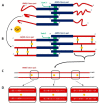
The schematic view of possible AMBN oligomerization state. AMBN monomers forming oligomers (e.g., hexamer). The blue rectangle illustrates AMBN-Nterm, while the green rectangle shows oligomerization inducing exon 5. Red lines show the possible arrangements of AMBN-Cterm in the absence of Ca2+ (A) and in the presence of Ca2+ (B) with yellow circles representing Ca2+. Black lines represent possible Ca2+ binding to AMBN-Cterm. (C) Schematic detail of possible Ca2+-binding sites of two C-termini of AMBN. Black rectangles represent sequences with a high abundance of negatively charged amino acids. (D) Detail view of potential Ca2+-binding sites at AMBN C-termini is shown in panel C. Amino acid sequences of AMBN-Cterm are in black rectangles. Negatively charged amino acids as a high preferable binding sites are bold white.

Similar articles

Cited by

References

    1. Wald T., Bednarova L., Osicka R., Pachl P., Sulc M., Lyngstadaas S.P., Slaby I., Vondrasek J. Biophysical characterization of recombinant human ameloblastin. Eur. J. Oral Sci. 2011;119:261–269. doi: 10.1111/j.1600-0722.2011.00913.x. - DOI - PubMed
    1. Wald T., Osickova A., Sulc M., Benada O., Semeradtova A., Rezabkova L., Veverka V., Bednarova L., Maly J., Macek P., et al. Intrinsically Disordered Enamel Matrix Protein Ameloblastin Forms Ribbon-like Supramolecular Structures via an N-terminal Segment Encoded by Exon 5. J. Biol. Chem. 2013;288:22333–22345. doi: 10.1074/jbc.M113.456012. - DOI - PMC - PubMed
    1. Stakkestad O., Lyngstadaas S.P., Thiede B., Vondrasek J., Skalhegg B.S., Reseland J.E. Phosphorylation Modulates Ameloblastin Self-assembly and Ca2+ Binding. Front. Physiol. 2017;8:10. doi: 10.3389/fphys.2017.00531. - DOI - PMC - PubMed
    1. Boskey A.L., Villarreal-Ramirez E. Intrinsically disordered proteins and biomineralization. Matrix Biol. 2016;52–54:43–59. doi: 10.1016/j.matbio.2016.01.007. - DOI - PMC - PubMed
    1. Kalmar L., Homola D., Varga G., Tompa P. Structural disorder in proteins brings order to crystal growth in biomineralization. Bone. 2012;51:528–534. doi: 10.1016/j.bone.2012.05.009. - DOI - PubMed

LinkOut - more resources