Review
. 2020 Dec 5;8(12):1933.
doi: 10.3390/microorganisms8121933.

The T3SS of Shigella: Expression, Structure, Function, and Role in Vacuole Escape

Affiliations
Review

The T3SS of Shigella: Expression, Structure, Function, and Role in Vacuole Escape

Waad Bajunaid et al. Microorganisms. .

Abstract

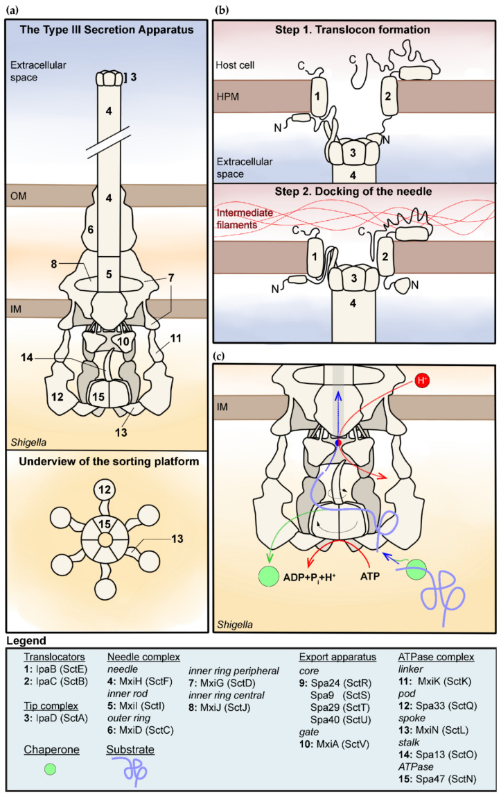
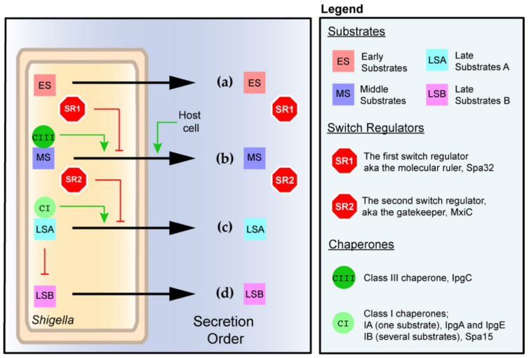
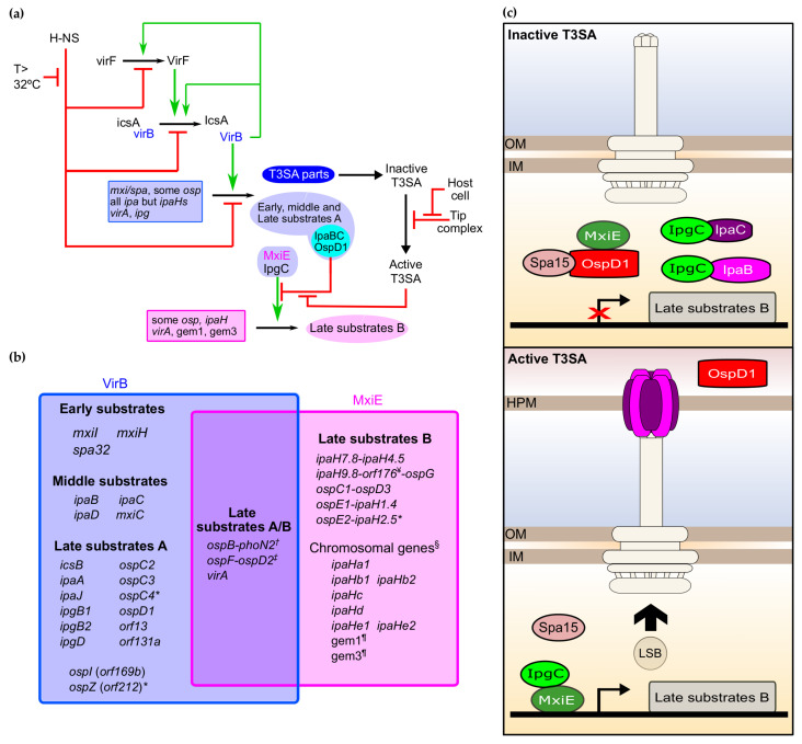
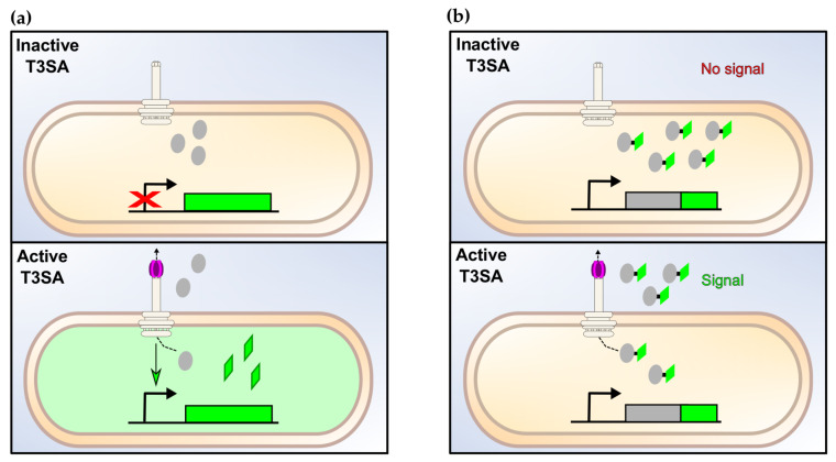
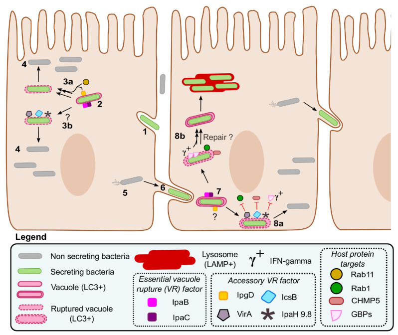
Shigella spp. are one of the leading causes of infectious diarrheal diseases. They are Escherichia coli pathovars that are characterized by the harboring of a large plasmid that encodes most virulence genes, including a type III secretion system (T3SS). The archetypal element of the T3SS is the injectisome, a syringe-like nanomachine composed of approximately 20 proteins, spanning both bacterial membranes and the cell wall, and topped with a needle. Upon contact of the tip of the needle with the plasma membrane, the injectisome secretes its protein substrates into host cells. Some of these substrates act as translocators or effectors whose functions are key to the invasion of the cytosol and the cell-to-cell spread characterizing the lifestyle of Shigella spp. Here, we review the structure, assembly, function, and methods to measure the activity of the injectisome with a focus on Shigella, but complemented with data from other T3SS if required. We also present the regulatory cascade that controls the expression of T3SS genes in Shigella. Finally, we describe the function of translocators and effectors during cell-to-cell spread, particularly during escape from the vacuole, a key element of Shigella's pathogenesis that has yet to reveal all of its secrets.

Keywords: Shigella; autophagy; genetically encoded reporter; injectisome; secretion; transcription regulation; type III secretion system (T3SS); vacuole rupture; virulence.


Conflict of interest statement

The authors declare no conflict of interest.

Figures

Figure 1
Figure 1
Structure and function of the type III secretion apparatus (T3SA) in Shigella spp. (a) Overview of the structure of the inactive T3SA. Note that the tip complex and cytosolic components of the T3SA are represented with a 3D perspective while the body is represented as a flat longitudinal cross section [16,17,31,32]. The bottom panel represents the sorting platform viewed from the cytosol of the bacterium. (i.e., viewed from the underside). (b) Model for the formation of the translocon and mutual interaction with the tip complex, the host plasma membrane, and the intermediate filaments [33,34]. (c) This is a model for the secretion of T3SA substrates summarizing elements discussed in the text. It indicates the role of the chaperone and ATPase–stalk complex in the unfolding of the substrates (purple) through rotation induced by ATP hydrolysis [35,36], and of the gate of the export apparatus in the creation of a proton motive force required for substrates secretion [37,38]. Movement is represented by dashed arrows; rotation is represented by curved arrows. The coupling of the protein substrates–proton antiporter is represented by the red–blue circle. The legend indicates the name of the various components in Shigella and in the unified nomenclature in parentheses.
Figure 2
Figure 2
The ordered secretion of the T3SA in Shigella and its main regulators. (a) The early substrates are secreted as soon as expressed to assemble the inner rod and needle. The concomitant secretion of the first switch regulator Spa32 allows secretion of small amounts of middle substrates IpaD and IpaB to yield the inactive T3SA represented in Figure 1a. (b) The contact with host cells activates the T3SA, allowing the secretion of middle substrates and the second switch regulator MxiC. This, in turn, triggers the successive secretion of late substrates A (c) and late substrates B (d). The black arrow represents the secretion by the T3SA. The control of the production of late substrates B by late substrates A is described in Figure 3.
Figure 3
Figure 3
The transcription regulatory cascade of the T3SA. (a) Key transcriptional regulatory cascade of the type III secretion system (T3SS) in Shigella. (b) The VirB and MxiE regulons; this figure is inspired from a prior version [95], and updated to take into account the most recent findings [58,96]. * These are likely pseudogenes in strain M90T; this gene belongs to the VirB and MxiE regulon but its product is not a T3SA substrate; this gene is weakly expressed and a new addition to the MxiE regulon; § the chromosomal ipaHs are annotated according to [97]; whether the product of these genes are T3SA substrates is currently unknown; ¥ this gene encodes an antitoxin from a TA system that is weakly secreted by the T3SA. Note that other antitoxin coding genes gmvA (orf48), orf86, and mvpA, which are not part of these two regulons, were also suggested to be T3SA substrates [58]. (c) Heteromers formed by MxiE and IpgC in the inactive and active state of the T3SA and their effect on the transcription of late substrates B.
Figure 4
Figure 4
The types of assays to measure the activity of the T3SA. (a) Transcription-based assays. The activation of T3SAs upregulate the expression of the reporter gene, thus leading to an increased production of the corresponding assay proteins. The relative secretion activity is then estimated by the measurement of the fluorescence or of any other relevant signal emitted by the assay protein (e.g., luminescence, colorimetric, etc.). (b) Secretion-based assay. Upon activation of the T3SA, T3SA substrates fused to a specific tag are secreted. The relative amount and location of the substrates inside host cells or in the extracellular medium are measured with the relevant method, as described in (a).
Figure 5
Figure 5
Shigella intracellular lifestyle and role of its virulence factors during vacuole escape. The invasion of a nonphagocytic host cell starts by the activation of T3SAs, which induce the remodeling of the plasma membrane through the action of their translocators and effectors (1). The intracellular bacterium is captured in a single membrane entry vacuole; several T3SA substrates, including the translocators IpaB, IpaC, and IpgD, accumulate in the vicinity of the vacuole (2). IpaBC form pores that are essential to vacuole rupture; the IpgD-dependent recruitment of Rab11 might accelerate this process (3a); although the other accessory vacuole rupture factors IcsB, VirA, and IpaH9.8 are dispensable during entry in epithelial cells, they might be important in yet undiscovered conditions such as in immune cells or in specific cytokinic contexts (3b). Cytosolic bacteria proliferate (4) and move using actin comets (5). Upon contact with the plasma membrane, the T3SA is reactivated, allowing the formation of a protrusion (6) and a double membrane dissemination vacuole (7). IpaB and IpaC initiate the rupture of the double membrane dissemination vacuole; the role of IpgD at this stage is unknown. The accessory VR factors IcsB and VirA, and probably IpaH9.8 in the presence of interferon-gamma (IFNγ), facilitate the escape of Shigella from the dissemination vacuole already partly ruptured by IpaBC (8a); this is probably realized through the inhibition of their respective targets CHMP5, Rab1, and GBPs. These WT bacteria can then resume cell-to-cell spread. By contrast, in the absence of these accessory VR factors, the repair of the partially ruptured vacuole is induced by Rab1 and CHMP5 (8b), a process that might also be facilitated by the action of the GBPs when IFNγ is present. These mutants (8b) are more often captured in lysosomes and partly deficient in cell-to-cell spread.

Similar articles

Cited by

References

    1. Kotloff K.L., Riddle M.S., Platts-Mills J.A., Pavlinac P., Zaidi A.K.M. Shigellosis. Lancet. 2018;391:801–812. doi: 10.1016/S0140-6736(17)33296-8. - DOI - PubMed
    1. Lampel K.A., Formal S.B., Maurelli A.T. A brief history of Shigella. EcoSal Plus. 2018;8:1–25. doi: 10.1128/ecosalplus.ESP-0006-2017. - DOI - PMC - PubMed
    1. Campbell-Valois F.-X., Pontier S.M. Implications of spatiotemporal regulation of Shigella flexneri type three secretion activity on effector functions: Think globally, act locally. Front. Cell. Infect. Microbiol. 2016;6:28. doi: 10.3389/fcimb.2016.00028. - DOI - PMC - PubMed
    1. Izoré T., Job V., Dessen A. Biogenesis, regulation, and targeting of the type III secretion system. Structure. 2011;19:603–612. doi: 10.1016/j.str.2011.03.015. - DOI - PubMed
    1. Deng W., Marshall N.C., Rowland J.L., McCoy J.M., Worrall L.J., Santos A.S., Strynadka N.C.J., Finlay B.B. Assembly, structure, function and regulation of type III secretion systems. Nat. Rev. Microbiol. 2017;15:323–337. doi: 10.1038/nrmicro.2017.20. - DOI - PubMed

Grants and funding

LinkOut - more resources