Case Reports
. 2020 Dec 4;7(3):57-63.
doi: 10.3390/dermatopathology7030010.

Type I Interferon Signature in Chilblain-Like Lesions Associated with the COVID-19 Pandemic

Affiliations
Case Reports

Type I Interferon Signature in Chilblain-Like Lesions Associated with the COVID-19 Pandemic

Roland Aschoff et al. Dermatopathology (Basel). .

Abstract

Contemporarily to the new SARS-CoV-2 mediated COVID-19 pandemic, a rise in patients with acral chilblain lesions has been described. They manifest late after mild disease or asymptomatic exposure to SARS-CoV-2. Their pathogenic evolution is currently unknown. In biopsies from three patients with acral partially ulcerating chilblain lesions that occurred associated to the COVID-19 pandemic, we analysed the expression of type I interferon induced proteins and signal transduction kinases. Histology demonstrated perivascular and periadnexal lymphohistiocytic infiltrates and endothelial dominated MxA-staining, as well as pJAK1 activation. Our findings demonstrate induction of the type I IFN pathway in lesional sections of COVID-19-associated chilblain-like lesions. This may indicate a local antiviral immune activation status associated with preceding exposure to SARS-CoV-2.

Keywords: SARS-CoV-2; chilblain; type I interferon.


Conflict of interest statement

The authors state no conflict of interest.

Figures

Figure 1
Figure 1
Clinical presentation of chilblain-like lesions on the feet and toes of three young men. Purple to red, tender chilblain-like lesions, superficial blisters and ulcerations on acral locations of the feet occurring 5 weeks after a travel through Germany in February 2020 in Patient 1 (A), 1 month after severe respiratory infection in Patient 2 (B), and 1 month after holidays in Tirol, Austria, that became a hotspot for COVID-19, in Patient 3 (C).
Figure 2
Figure 2
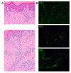
Histological pattern of chilblain-like lesions. Hematoxylin Eosin (HE) staining of chilblain-like lesions demonstrating superficial and deep perivascular and periadnexal lymphocytic infiltrates (A), magnification ×200 and ×100. Immunofluorescence staining shows deposition of c3, IgG and IgM in dermal vessels and granular c3 and IgM deposits at the basement membrane zone (B), magnification ×400. Histology of Patient 2 is shown as representative of similar results in all three patients.
Figure 3
Figure 3
Immunohistochemistry demonstrating type I IFN activation. (AD): Staining for MxA in red is negative in healthy skin (A), shows strong staining in conventional chilblain lupus (B), and protein induction in epidermis, dermis and endothelial cells (arrow) in lesional skin of chilblain-like lesions after exposure to SARS-CoV-2 (C,D arrow). (EH): Staining for pJAK1 in red is negative in healthy skin (E), shows faint staining in conventional chilblain lupus (F), and staining in the epidermis, dermis and endothelial cells (arrow) in lesional skin of chilblain-like lesions after exposure to SARS-CoV-2 (G,H). (IL): Staining for pJAK2 in red is negative in healthy skin (I), shows staining in chilblain lupus (J), and faint staining in the epidermis, dermis and endothelial cells (arrow) in lesional skin of chilblain-like lesions after exposure to SARS-CoV-2 (K,L). (C,D,G,H,K,L) show representative images of stainings performed in all 3 patients. D, H, L show magnifications of the respective staining in (C,G,K).

References

    1. Mehra M.R., Desai S.S., Kuy S., Henry T.D., Patel A.N. Cardiovascular Disease, Drug Therapy, and Mortality in Covid-19. N. Engl. J. Med. 2020;382:e102. doi: 10.1056/NEJMoa2007621. - DOI - PMC - PubMed
    1. Gunther C., Aschoff R., Beissert S. Cutaneous autoimmune diseases during COVID-19 pandemic. J. Eur. Acad. Dermatol. Venereol. 2020 doi: 10.1111/jdv.16753. - DOI - PMC - PubMed
    1. Fernandez-Nieto D., Jimenez-Cauhe J., Suarez-Valle A., Moreno-Arrones O.M., Saceda-Corralo D., Arana-Raja A., Ortega-Quijano D. Characterization of acute acral skin lesions in nonhospitalized patients: A case series of 132 patients during the COVID-19 outbreak. J. Am. Acad. Dermatol. 2020;83:e61–e63. doi: 10.1016/j.jaad.2020.04.093. - DOI - PMC - PubMed
    1. Landa N., Mendieta-Eckert M., Fonda-Pascual P., Aguirre T. Chilblain-like lesions on feet and hands during the COVID-19 Pandemic. Int. J. Dermatol. 2020;59:739–743. doi: 10.1111/ijd.14937. - DOI - PMC - PubMed
    1. Piccolo V., Neri I., Filippeschi C., Oranges T., Argenziano G., Battarra V.C., Berti S., Manunza F., Fortina A.B., Di Lernia V., et al. Chilblain-like lesions during COVID-19 epidemic: A preliminary study on 63 patients. J. Eur. Acad. Dermatol. Venereol. 2020;34:e291–e293. doi: 10.1111/jdv.16526. - DOI - PMC - PubMed

Publication types

LinkOut - more resources