. 2020 Dec 4;12(12):3640.
doi: 10.3390/cancers12123640.

Growth Hormone Upregulates Mediators of Melanoma Drug Efflux and Epithelial-to-Mesenchymal Transition In Vitro and In Vivo

Affiliations

Growth Hormone Upregulates Mediators of Melanoma Drug Efflux and Epithelial-to-Mesenchymal Transition In Vitro and In Vivo

Yanrong Qian et al. Cancers (Basel). .

Abstract

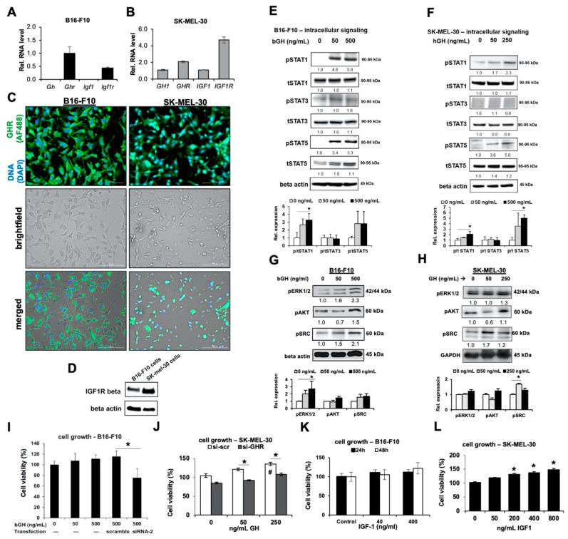
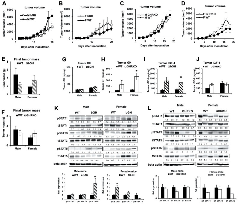
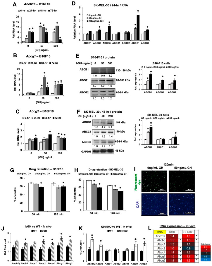
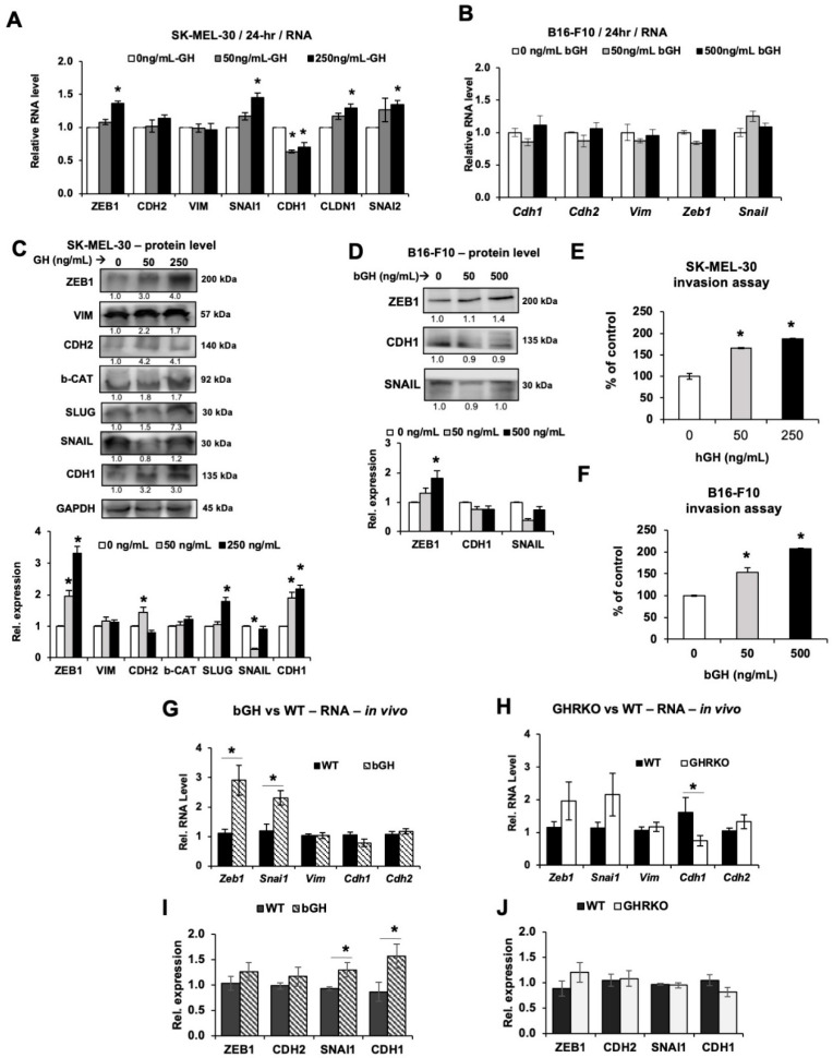
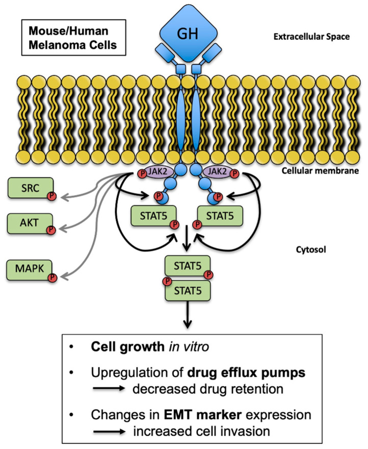
Growth hormone (GH) and the GH receptor (GHR) are expressed in a wide range of malignant tumors including melanoma. However, the effect of GH/insulin-like growth factor (IGF) on melanoma in vivo has not yet been elucidated. Here we assessed the physical and molecular effects of GH on mouse melanoma B16-F10 and human melanoma SK-MEL-30 cells in vitro. We then corroborated these observations with syngeneic B16-F10 tumors in two mouse lines with different levels of GH/IGF: bovine GH transgenic mice (bGH; high GH, high IGF-1) and GHR gene-disrupted or knockout mice (GHRKO; high GH, low IGF-1). In vitro, GH treatment enhanced mouse and human melanoma cell growth, drug retention and cell invasion. While the in vivo tumor size was unaffected in both bGH and GHRKO mouse lines, multiple drug-efflux pumps were up regulated. This intrinsic capacity of therapy resistance appears to be GH dependent. Additionally, epithelial-to-mesenchymal transition (EMT) gene transcription markers were significantly upregulated in vivo supporting our current and recent in vitro observations. These syngeneic mouse melanoma models of differential GH/IGF action can be valuable tools in screening for therapeutic options where lowering GH/IGF-1 action is important.

Keywords: epithelial-to-mesenchymal transition; growth hormone; growth hormone receptor; insulin-like growth factor-1; melanoma; multidrug efflux pumps.


Conflict of interest statement

The authors declare no conflict of interest.

Figures

Figure 1
Figure 1
Melanoma cells are responsive to GH treatment in vitro. (A) Relative RNA expression was quantified for Gh, Ghr, Igf1 and Igf1r genes in B16-F10 mouse melanoma cells (n = 3). (B) Relative RNA expression was quantified for GH, GHR, IGF1 and IGF1R genes in SK-MEL-30 human melanoma cells, normalized against GAPDH (n = 3). (C) B16-F10 and SK-MEL-30 cells were stained with GHR antibody. Top panels present cellular DNA stained with DAPI (blue) and fluorescent signals from AF488-tagged anti-GHR antibody (green). Middle panels represent brightfield views while bottom panels represent merged views (20× magnification; scale bar represents 100 μm). (D) Expression of IGF1R-beta was detected in B16-F10 and SK-MEL-30 cells via western blotting. (E) Representative images of western blot analyses of phosphorylation (p) and total (t) levels of STATs 1, 3 and 5 in bovine GH (bGH) treated B16-F10 melanoma cell lysates. Cells were treated for 30 min and lysed. Densitometry analysis was performed and normalized against β-actin. The fold changes relative to control are labeled under each band. The ratio of p/t protein levels are quantified against controls and shown in bar graphs (n = 3). (F) Similar results for SK-MEL-30 cells treated with human (h) GH for 30 min were obtained. (G,H) Changes in GH signaling proteins, pERK1/2, pAKT and pSRC, in B16-F10 cells and SK-MEL-30 cells treated with bGH or hGH for 48 h. Protein levels were normalized against β-actin or GAPDH. (I) Changes in cell proliferation of B16-F10 cells transfected with scramble or GHR-targeted siRNA (siRNA-2), after 48-h treatment with bGH. (J) Similar results were observed in SK-MEL-30 cells transfected with scramble (si-scr) or GHR-targeted siRNA (si-GHR), after 48-h treatment with hGH (* as compared with si-scr group, # as compared with no treatment group). (K) Changes in cell proliferation of B16-F10 cells treated with increasing doses of IGF-1 for 24 and 48 h. (L) Changes in cell proliferation of SK-MEL-30 after 48-h treatment with recombinant human IGF-1 (n = 5). Data are presented as mean ± standard deviation (*, p < 0.05, Mann-Whitney U test).
Figure 2
Figure 2
Subcutaneous B16-F10 mouse melanoma growth in bGH and GHRKO syngeneic mice. (A) Subcutaneous B16-F10 tumor growth in male bGH (n = 5) vs. WT mice (n = 6). (B) Subcutaneous B16-F10 tumor growth in female bGH (n = 7) and WT mice (n = 6). (C) Subcutaneous B16-F10 tumor growth in male GHRKO (n = 6) vs. WT (n = 8) mice. (D) Subcutaneous B16-F10 tumor growth in female GHRKO (n = 4) and WT mice (n = 5). Tumor sizes were analyzed by repeated measures (SPSS). (E) Weight of subcutaneous B16-F10 tumors from bGH vs. WT mice at dissection. (F) Weight of subcutaneous B16-F10 tumors from GHRKO vs. WT mice at dissection. (G) GH levels were measured in protein lysates isolated from tumors of bGH and WT mice using ELISA and normalized to total protein concentrations (n = 4). (H) Similar GH measurements were performed in protein lysates isolated from tumors of GHRKO and WT mice (males n = 3, females n = 4). (I) IGF-1 levels were measured in protein lysates isolated from tumors of bGH and WT mice using ELISA and normalized to total protein concentrations (n = 4). (J) Similar IGF-1 measurements were performed in protein lysates isolated from tumors of GHRKO and WT mice (males n = 3, females n = 4). (K) Representative images of western blot analysis of phosphorylation (p) and total (t) levels of STAT1, STAT3 and STAT5 in protein lysates isolated from tumors of bGH and WT mice. Densitometry analysis was performed and normalized against β-Actin. The relative expression levels (fold change relative to WT) are labeled under each band. The ratio of phosphorylated vs. total protein levels in tumors from bGH and WT mice are presented in bar graphs (n = 4). (L) Representative images of western blot analysis of phosphorylation and total levels of STAT1, STAT3, and STAT5 in protein lysates isolated from tumors of GHRKO and WT mice. (males n = 3, females n = 4). Data are presented as mean ± standard errors (*, p < 0.05, unpaired student’s t-test).
Figure 3
Figure 3
Changes in ABC efflux pumps in B16-F10 mouse melanoma in vitro and in vivo. (AC) Changes in RNA levels for Abcb1a, Abcg1, Abcg2 in B16-F10 mouse melanoma cells treated with bGH, normalized against reference genes (n = 3). (D) Changes in RNA levels for several ABC efflux pumps in SK-MEL-30 human melanoma cells treated with hGH for 24 h, normalized against GAPDH gene (n = 3). (E) Changes in proteins levels of several ABC efflux pumps of B16-F10 cells treated with bGH for 72 h. Densitometry analysis was performed and normalized to total protein (n = 3). (F) Changes in proteins levels of several ABC efflux pumps of SK-MEL-30 cells treated with hGH for 48 h, normalized to GAPDH (n = 3). (G) Drug retention was decreased in B16-F10 cells pretreated with bGH for 1 week (n = 3). (H) Similar results were found in SK-MEL-30 cells (n = 3). Data are presented as mean ± standard deviation (*, p < 0.05, Mann-Whitney U test). (I) Representative images for SK-MEL-30 cells with fluorescent dye (green) treated with and without 50 ng/mL hGH for 1 week (20× magnification; scale bar represents 300 μm). (J) Relative RNA levels for seven different ABC efflux pumps in B16-F10 tumors grown in vivo in bGH and WT mice, normalized against reference genes (both sexes combined). bGH mice (n = 12), WT mice (n = 12). (K) Relative RNA levels for seven different ABC efflux pumps in B16-F10 tumors grown in vivo in GHRKO and WT mice, normalized against reference genes (both sexes combined). GHRKO mice (n = 10), WT mice (n = 13). (L) Heatmap showing the variations in RNA expression of ABC efflux pumps in tumors in bGH or GHRKO mice (both sexes combined). Data are presented as mean ± standard error (*, p < 0.05, unpaired student’s t-test).
Figure 4
Figure 4
Changes in markers of epithelial-to-mesenchymal transition (EMT) in melanoma in vitro and in vivo. (A) Changes in RNA levels in SK-MEL-30 cells treated with hGH for 24 h, normalized against GAPDH gene. (B) Relative RNA levels in B16-F10 cells treated with bGH for 24 h, normalized against reference genes. (C) Proteins levels of EMT markers in SK-MEL-30 cells after hGH treatment for 48 h, normalized to GAPDH. (D) Proteins levels of EMT markers in B16-F10 cells after bGH treatment for 72 h, normalized against total protein. (E,F) Invasion of SK-MEL-30 (E) and B16-F10 (F) cells after 7-days hGH or bGH pretreatment. Fold changes are presented and refer to control. Data are presented as mean ± standard deviation (n = 3; *, p < 0.05, Mann-Whitney U test). (G,H) Relative RNA levels of EMT markers in B16-F10 tumors grown in vivo in bGH (G) or GHRKO (H) mice, normalized against reference genes (both sexes combined). (I,J) Proteins levels of EMT markers in tumors from WT, bGH (I) or GHRKO (J) mice. Representative images are shown in Figure S6F,G. (both sexes combined; bGH mice n = 12, WT mice n = 12; GHRKO mice n = 10, WT mice n = 13). Data are presented as mean ± standard error (*, p < 0.05, unpaired student’s t-test).
Figure 5
Figure 5
Proposed model for this study. GH stimulates melanoma growth in vitro through GHR activation independent of IGF-1. In the syngeneic mouse melanoma models, elevated GH, independent of IGF-1, alters tumor expression of multidrug efflux pumps and epithelial-mesenchymal transition markers in vivo.

References

    1. Ikeno Y., Hubbard G.B., Lee S., Cortez L.A., Lew C.M., Webb C.R., Berryman D.E., List E.O., Kopchick J.J., Bartke A. Reduced incidence and delayed occurrence of fatal neoplastic diseases in growth hormone receptor/binding protein knockout mice. J. Gerontol. A Biol. Sci. Med. Sci. 2009;64:522–529. doi: 10.1093/gerona/glp017. - DOI - PMC - PubMed
    1. Pollak M., Blouin M.J., Zhang J.C., Kopchick J.J. Reduced mammary gland carcinogenesis in transgenic mice expressing a growth hormone antagonist. Br. J. Cancer. 2001;85:428–430. doi: 10.1054/bjoc.2001.1895. - DOI - PMC - PubMed
    1. Conway-Campbell B.L., Wooh J.W., Brooks A.J., Gordon D., Brown R.J., Lichanska A.M., Chin H.S., Barton C.L., Boyle G.M., Parsons P.G., et al. Nuclear targeting of the growth hormone receptor results in dysregulation of cell proliferation and tumorigenesis. Proc. Natl. Acad. Sci. USA. 2007;104:13331–13336. doi: 10.1073/pnas.0600181104. - DOI - PMC - PubMed
    1. Chien C.H., Lee M.J., Liou H.C., Liou H.H., Fu W.M. Growth hormone is increased in the lungs and enhances experimental lung metastasis of melanoma in DJ-1 KO mice. BMC Cancer. 2016;16 doi: 10.1186/s12885-016-2898-5. - DOI - PMC - PubMed
    1. Van der Lely A.J., Muller A., Janssen J.A., Davis R.J., Zib K.A., Scarlett J.A., Lamberts S.W. Control of tumor size and disease activity during cotreatment with octreotide and the growth hormone receptor antagonist pegvisomant in an acromegalic patient. J. Clin. Endocrinol. Metab. 2001;86:478–481. doi: 10.1210/jcem.86.2.7206. - DOI - PubMed

LinkOut - more resources