

Exosomes: Emerging Diagnostic and Therapeutic Targets in Cutaneous Diseases
- PMID: 33291683
- PMCID: PMC7730213
- DOI: 10.3390/ijms21239264
Exosomes: Emerging Diagnostic and Therapeutic Targets in Cutaneous Diseases
Abstract
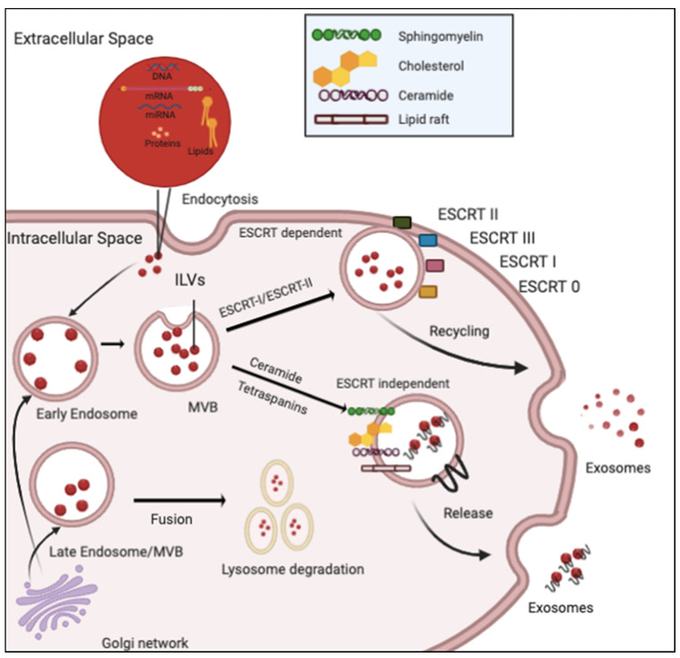
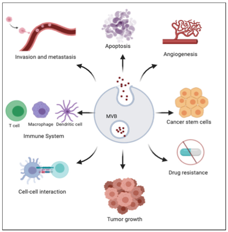
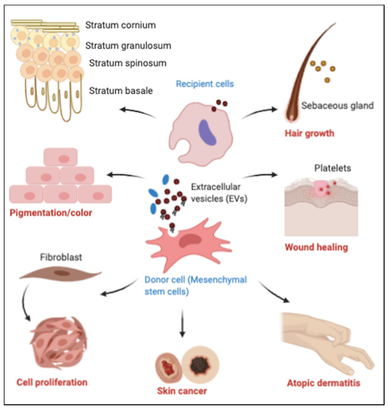
Skin is the largest human organ and is continuously exposed to various exogenous and endogenous trigger factors affecting body homeostasis. A number of mechanisms, including genetic, inflammatory and autoimmune ones, have been implicated in the pathogenesis of cutaneous diseases. Recently, there has been considerable interest in the role that extracellular vesicles, particularly exosomes, play in human diseases, through their modulation of multiple signaling pathways. Exosomes are nano-sized vesicles secreted by all cell types. They function as cargo carriers shuttling proteins, nucleic acids, lipids etc., thus impacting the cell-cell communications and transfer of vital information/moieties critical for skin homeostasis and disease pathogenesis. This review summarizes the available knowledge on how exosomes affect pathogenesis of cutaneous diseases, and highlights their potential as future targets for the therapy of various skin diseases.
Keywords: cancer; exosomes; extracellular vesicles; inflammation; skin.
Conflict of interest statement
The authors declare no conflict of interest.
Figures
References
-
- Karimkhani C., Dellavalle R.P., Coffeng L.E., Flohr C., Hay R.J., Langan S.M., Nsoesie E.O., Ferrari A.J., Erskine H.E., Silverberg J.I., et al. Global skin disease morbidity and mortality: An update from the global burden of disease study 2013. JAMA Dermatol. 2017;153:406–412. doi: 10.1001/jamadermatol.2016.5538. - DOI - PMC - PubMed
-
- Thery C., Witwer K.W., Aikawa E., Alcaraz M.J., Anderson J.D., Andriantsitohaina R., Antoniou A., Arab T., Archer F., Atkin-Smith G.K., et al. Minimal information for studies of extracellular vesicles 2018 (misev2018): A position statement of the international society for extracellular vesicles and update of the misev2014 guidelines. J. Extracell Vesicles. 2018;7:1535750. doi: 10.1080/20013078.2018.1535750. - DOI - PMC - PubMed
-
- Tello-Flores V.A., Valladares-Salgado A., Ramirez-Vargas M.A., Cruz M., Del-Moral-Hernandez O., Cahua-Pablo J.A., Ramirez M., Hernandez-Sotelo D., Armenta-Solis A., Flores-Alfaro E. Altered levels of malat1 and h19 derived from serum or serum exosomes associated with type-2 diabetes. Noncoding RNA Res. 2020;5:71–76. doi: 10.1016/j.ncrna.2020.03.001. - DOI - PMC - PubMed
Publication types
MeSH terms
Substances
LinkOut - more resources
Full Text Sources
Other Literature Sources
Medical
