Review
. 2020 Dec 4;9(12):2604.
doi: 10.3390/cells9122604.

TRPM Channels in Human Diseases

Affiliations
Review

TRPM Channels in Human Diseases

Ivanka Jimenez et al. Cells. .

Abstract

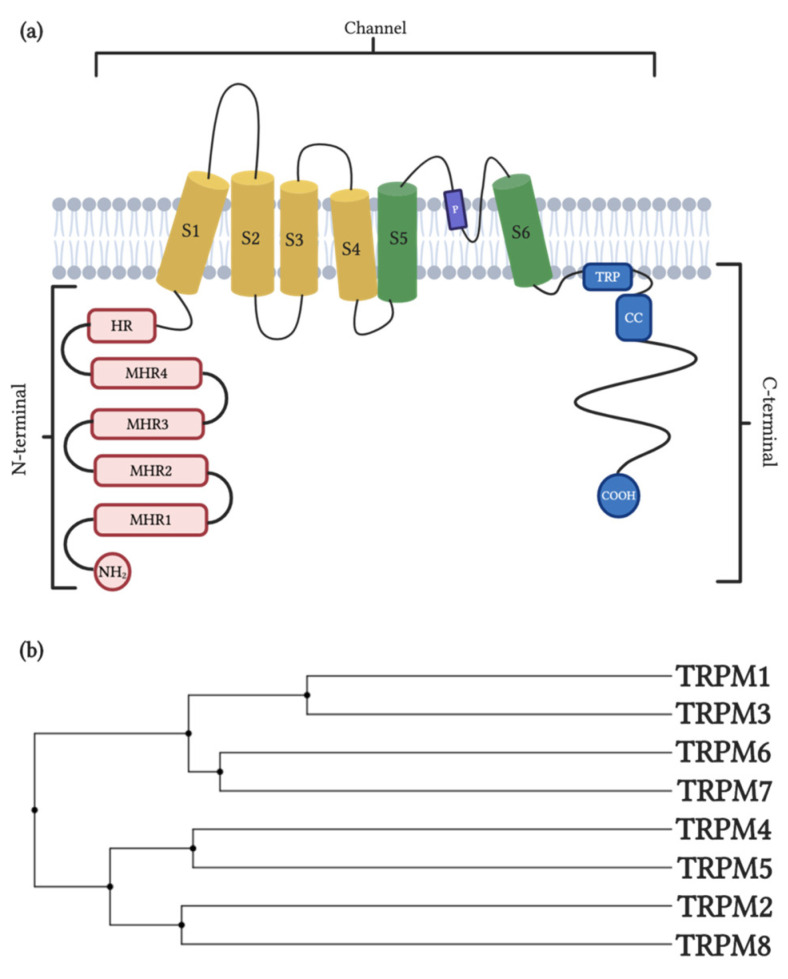
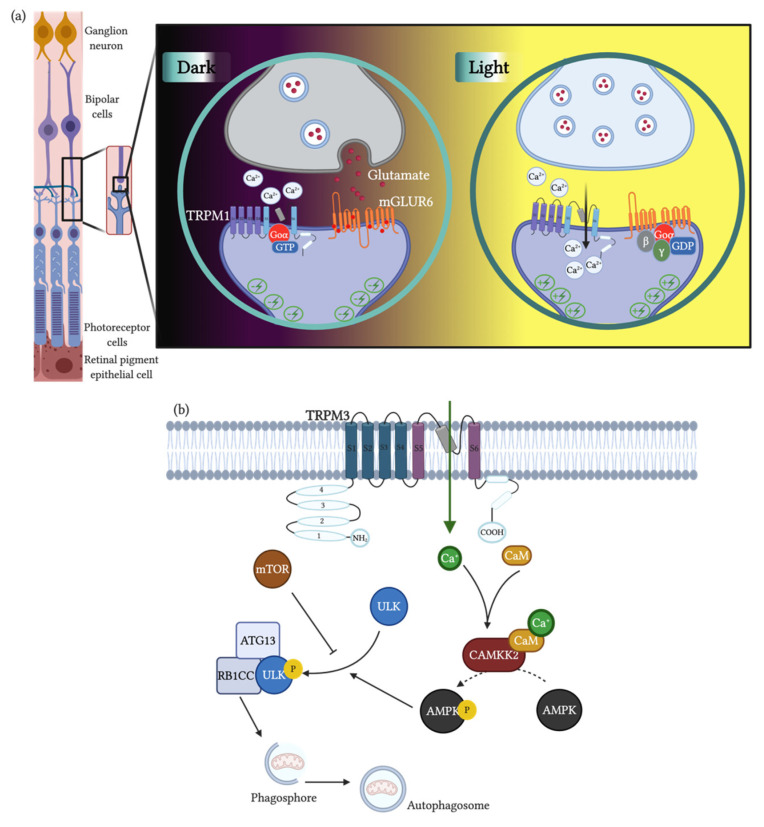
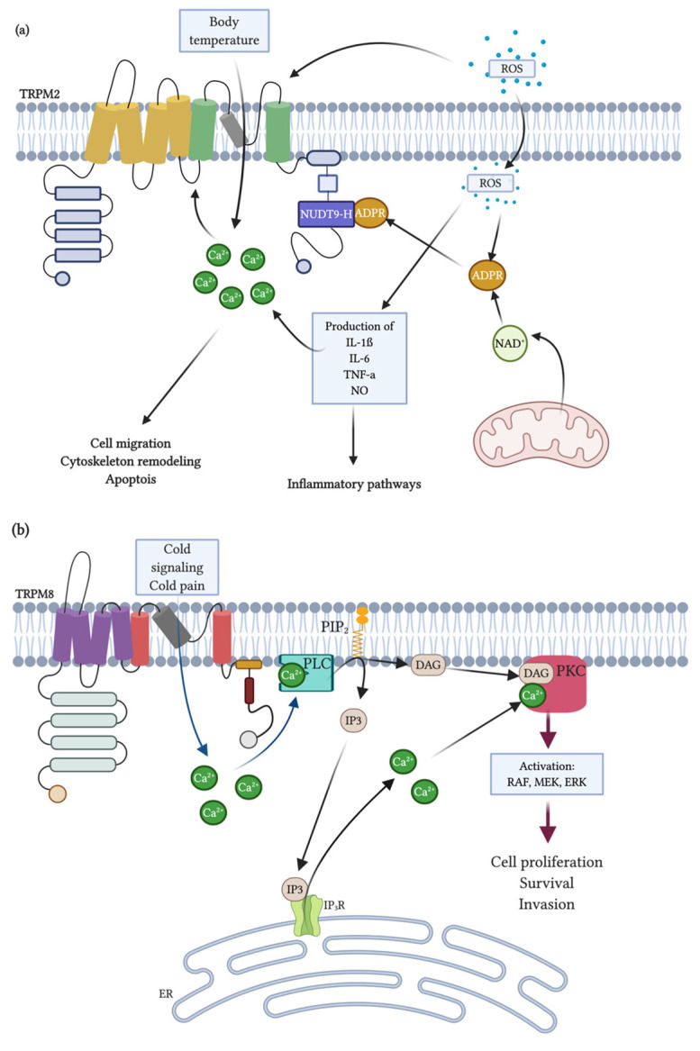
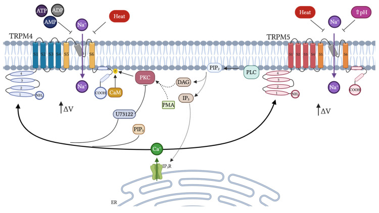
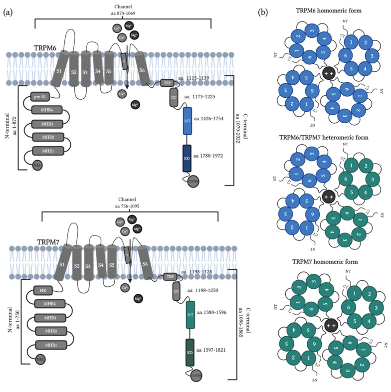
The transient receptor potential melastatin (TRPM) subfamily belongs to the TRP cation channels family. Since the first cloning of TRPM1 in 1989, tremendous progress has been made in identifying novel members of the TRPM subfamily and their functions. The TRPM subfamily is composed of eight members consisting of four six-transmembrane domain subunits, resulting in homomeric or heteromeric channels. From a structural point of view, based on the homology sequence of the coiled-coil in the C-terminus, the eight TRPM members are clustered into four groups: TRPM1/M3, M2/M8, M4/M5 and M6/M7. TRPM subfamily members have been involved in several physiological functions. However, they are also linked to diverse pathophysiological human processes. Alterations in the expression and function of TRPM subfamily ion channels might generate several human diseases including cardiovascular and neurodegenerative alterations, organ dysfunction, cancer and many other channelopathies. These effects position them as remarkable putative targets for novel diagnostic strategies, drug design and therapeutic approaches. Here, we review the current knowledge about the main characteristics of all members of the TRPM family, focusing on their actions in human diseases.

Keywords: TRPM channels; human diseases; ion channels.


Conflict of interest statement

The authors declare no conflict of interest.

Figures

Figure 1
Figure 1
Channel structure and family tree of the transient receptor potential melastatin (TRPM) subfamily. (a) The N-terminus is composed of four melastatin homology regions and homology region pre-S1 (melastatin homology regions (MHR) and homology regions (HR), red boxes). The channel domain contains six transmembrane segments (S1–S6), S1–S4 (yellow cylinder) corresponding to a voltage-sensor-like domain; the pore is formed by the loop between the S5 and S6 segments (purple box and green cylinder). The C-terminus is composed of TRP and the coiled-coil (CC) (blue boxes). (b) Phylogeny of human TRPM channels. Created with BioRender.com.
Figure 2
Figure 2
TRPM1 role in visual pathways. In the retina, the photoreceptors are in synaptic contact with bipolar neurons. (a), which in turn contact ganglion neurons, in which axons merge to form the optic nerve. In the dark, the rods secrete glutamate into the synaptic cleft, which activates the mGluR6, a G-protein coupled receptor. In its activated state, mGluR6 hyperpolarizes the neuron. In that status, G-protein Goα-GTP is bound to TRPM1, resulting in inactivation of the channel. In the presence of light, rods release low glutamate, which inactivates mGluR6. As a consequence, the dephosphorylation of GTP into GDP allows Go to bind and subunits and the complex binds mGluR6, inactivating it and avoiding hyperpolarization. The release of Go activates TRPM1, which opens, allowing calcium entry and depolarizing the bipolar neuron triggering the electrochemical signaling. (b) TRPM3 and autophagy in clear cell carcinoma. TRPM3 activity increases cytosolic calcium, which binds Calmodulin (CaM). Ca-CaM binds CaMKK2 and this activated complex phosphorylates AMPK. Active AMPK through phosphorylation, in turn, phosphorylates ULK, which, in its phosphorylated form, can bind ATG13 and RB1CC1. This ULK-ATG13-RB1CC1 complex allows for the formation of autophagosomes and the binding of the autophagosomes with lysosomes. ULK is normally inactive through the phosphatase activity of MTOR. Thus, in normal conditions, this pathway is inactive. Created with BioRender.com.
Figure 3
Figure 3
Signaling mechanisms for TRPM2 and TRPM8 activation. (a) NAD+ and reactive oxygen species (ROS), including H2O2, accumulate during inflammation and tissue damage may trigger TRPM2. NAD+ may be converted to ADPR and cADPR. ROS can also cross the plasma membrane and mobilize ADPR from mitochondria and both ROS and cADPR can synergize with ADPR to activate TRPM2. Additionally, ADPR is generated from NAD+ via poly-ADPR during ROS-induced damage through the activation of PARP (poly(ADP-ribose) polymerase)-ADPR-dependent mechanisms in inflammatory cells. Free cytosolic ADPR can act on the NUDT9-H of TRPM2 channels, enabling Ca2+ influx across the plasma membrane and/or release of intracellular Ca2+, raising the Ca2+ concentration in the cytosol. On the other hand, ROS induces cytokine production (pro-inflammatory response), which may alter intracellular calcium levels. A Ca2+ increase will activate different physiological processes including gene expression through Ca2+-dependent signaling pathways such as apoptosis, cell migration and cytoskeleton remodeling. Moreover, TRPM2 may detect increased temperatures to prevent overheating, limiting the fever response. (b) Cold temperature, menthol or icilin can stimulate the activity of the TRPM8 ion channels. Upon activation, the TRPM8 channel changes conformation and permits extracellular Ca2+ to flow through it across membranes. This results in the activation of Ca2+-sensitive PLC and the hydrolysis of PIP2, which produces inositol 1,4,5-triphosphate (IP3) that causes the release of Ca2+ from the intracellular stores and generates diacylglycerol (DAG). The elevated Ca2+ level or DAG can activate protein kinase C (PKC), which, in turn, activates RAF in the ERK pathway. Therefore, transcriptional activation of genes stimulates cellular proliferation, survival and invasion, which contribute to cancer growth and metastasis. Created with BioRender.com.
Figure 4
Figure 4
Endogenous regulation of TRPM4 and TRPM5. Both TRPM4 and TRPM5 are activated by [Ca2+]i and regulated by PIP2. TRPM4 also is activated by CaM and PKC by phosphorylation of the TRP region. PLC is fundamental for the regulation of both channels due to the activation of PKC via DAG and also mediates the release of calcium from ER via IP3. Finally, these channels are inhibited very differently. In the case of TRPM4, it is inhibited by AMP, ADP and ATP, while TRPM5 is inhibited by a high pH or heat. Created with BioRender.com.
Figure 5
Figure 5
TRPM6 and TRPM7 structure and tetramer representation. (a) The N-terminus region is composed of four melastatin homology regions (MHR) and the pre-S1 (TRPM6) or homology region (HR, TRPM7) domain. The channel domain contains six transmembrane segments (S1–S6) and the pore-forming loop between the S5 and S6 segments. The C-terminus region is composed of TRP, coiled-coil (CC), Serine—Threonine (S/T) and kinase domains. aa: amino acid. (b) These channels form tetramers with four subunits in homomeric or heteromeric form forming a pore in the membrane and allows divalent cations to permeate. 1–6: transmembrane subunits, NT: N-terminus, CT: C-terminus. Created with BioRender.com.

References

    1. Hübner C.A., Jentsch T.J. Ion channel diseases. Hum. Mol. Genet. 2002;11:2436–2447. doi: 10.1093/hmg/11.20.2435. - DOI - PubMed
    1. Niemeyer B.A., Mery L., Zawar C., Suckow A., Monje F., Pardo L.A., Stühmer W., Flockerzi V., Hoth M. Ion channels in health and disease. EMBO Rep. 2001;2:568–573. doi: 10.1093/embo-reports/kve145. - DOI - PMC - PubMed
    1. Hutchings C.J., Colussi P., Clark T.G. Ion channels as therapeutic antibody targets. MAbs. 2019;11:265–296. doi: 10.1080/19420862.2018.1548232. - DOI - PMC - PubMed
    1. Varela D., Simon F., Riveros A., Jørgensen F., Stutzin A. NAD(P)H oxidase-derived H2O2 signals chloride channel activation in cell volume regulation and cell proliferation. J. Biol. Chem. 2004;279:13301–13304. doi: 10.1074/jbc.C400020200. - DOI - PubMed
    1. Simon F., Varela D., Luisa Eguiguren A., Díaz L.F., Sala F., Stutzin A., Stutzin Hydroxyl A. Hydroxyl radical activation of a Ca2+-sensitive nonselective cation channel involved in epithelial cell necrosis. Am. J. Physiol. 2004;287:963–970. doi: 10.1152/ajpcell.00041.2004. - DOI - PubMed

Publication types