

Fabrication of Anisotropic Cu Ferrite-Polymer Core-Shell Nanoparticles for Photodynamic Ablation of Cervical Cancer Cells
- PMID: 33291730
- PMCID: PMC7761902
- DOI: 10.3390/nano10122429
Fabrication of Anisotropic Cu Ferrite-Polymer Core-Shell Nanoparticles for Photodynamic Ablation of Cervical Cancer Cells
Abstract
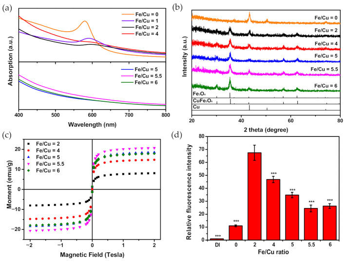
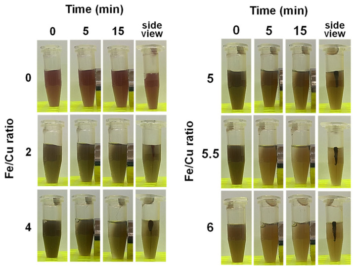
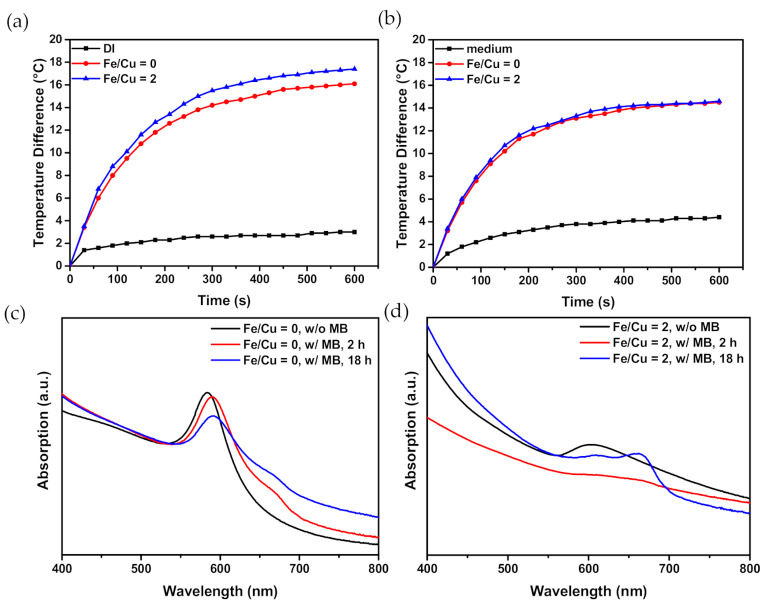
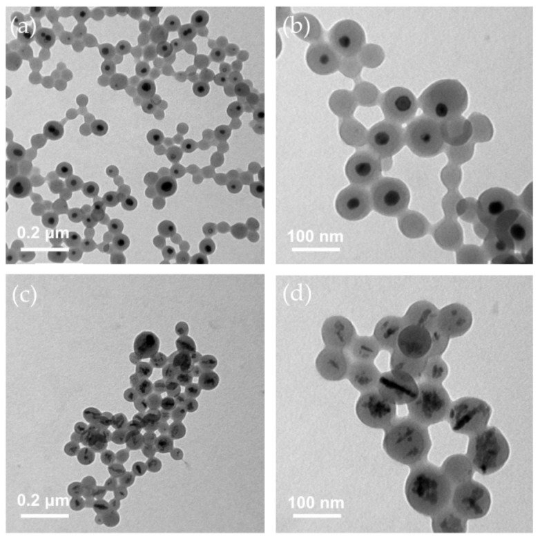
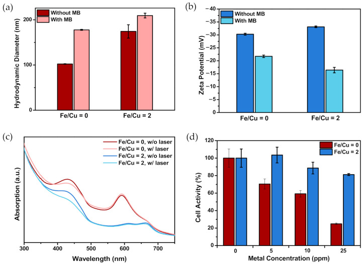
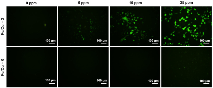
In this work we developed methylene blue-immobilized copper-iron nanoparticles (MB-CuFe NPs) through a facile one-step hydrothermal reaction to achieve a better phototherapeutic effect. The Fe/Cu ratio of the CuFe NPs was controllable by merely changing the loading amount of iron precursor concentration. The CuFe NPs could serve as a Fenton catalyst to convert hydrogen peroxide (H2O2) into reactive oxygen species (ROS), while the superparamagnetic properties also suggest magnetic resonance imaging (MRI) potential. Furthermore, the Food and Drug Administration (FDA)-approved MB photosensitizer could strongly adsorb onto the surface of CuFe NPs to facilitate the drug delivery into cells and improve the photodynamic therapy at 660 nm via significant generation of singlet oxygen species, leading to enhanced cancer cell-damaging efficacy. An MTT (thiazolyl blue tetrazolium bromide) assay proved the low cytotoxicity of the CuFe NPs to cervical cancer cells (HeLa cells), namely above 80% at 25 ppm of the sample dose. A slight dissolution of Cu and Fe ions from the CuFe NPs in an acidic environment was obtained, providing direct evidence for CuFe NPs being degradable without the risk of long-term retention in the body. Moreover, the tremendous photo-to-thermal conversion of CuFe NPs was examined, which might be combined with photodynamic therapy (PDT) for promising development in the depletion of cancer cells after a single pulse of deep-red light irradiation at high laser power.
Keywords: Fenton reaction; bimetallic nanoparticles; cancer treatment; photodynamic therapy; reactive oxygen species; superparamagnetic nanoparticles.
Conflict of interest statement
The authors declare no conflict of interest.
Figures

References
-
- Liu T.-M., Conde J., Lipiński T., Bednarkiewicz A., Huang C.-C. Revisiting the classification of NIR-absorbing/emitting nanomaterials for in vivo bioapplications. NPG Asia Mater. 2016;8:e295. doi: 10.1038/am.2016.106. - DOI
-
- Ahmadivand A., Gerislioglu B., Ahuja R., Mishra Y.K. Terahertz plasmonics: The rise of toroidal metadevices towards immunobiosensings. Mater. Today. 2020;32:108–130. doi: 10.1016/j.mattod.2019.08.002. - DOI
Grants and funding
LinkOut - more resources
Full Text Sources
Miscellaneous
