. 2020 Dec 28;60(12):6612-6623.
doi: 10.1021/acs.jcim.0c00877. Epub 2020 Dec 8.

Benchmark Sets for Binding Hot Spot Identification in Fragment-Based Ligand Discovery

Affiliations

Benchmark Sets for Binding Hot Spot Identification in Fragment-Based Ligand Discovery

Amanda E Wakefield et al. J Chem Inf Model. .

Abstract

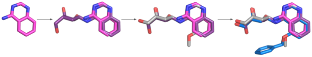
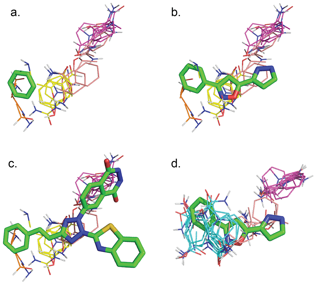
Binding hot spots are regions of proteins that, due to their potentially high contribution to the binding free energy, have high propensity to bind small molecules. We present benchmark sets for testing computational methods for the identification of binding hot spots with emphasis on fragment-based ligand discovery. Each protein structure in the set binds a fragment, which is extended into larger ligands in other structures without substantial change in its binding mode. Structures of the same proteins without any bound ligand are also collected to form an unbound benchmark. We also discuss a set developed by Astex Pharmaceuticals for the validation of hot and warm spots for fragment binding. The set is based on the assumption that a fragment that occurs in diverse ligands in the same subpocket identifies a binding hot spot. Since this set includes only ligand-bound proteins, we added a set with unbound structures. All four sets were tested using FTMap, a computational analogue of fragment screening experiments to form a baseline for testing other prediction methods, and differences among the sets are discussed.


Conflict of interest statement

Conflict of Interest Disclosure:

Acpharis Inc. offers commercial licenses to ATLAS, a software product in function similar to FTMap. Dima Kozakov and Sandor Vajda owns stock in the company. However, the FTMap program and the use of the FTMap server are free for academic and governmental use.

Figures

Figure 1.
Figure 1.
Chemical structures and PDB ligand ID codes of the fragments in the newly created Acpharis benchmark set.
Figure 2.
Figure 2.
Fragment 1LQ and ligands that contain the fragment as a substructure. The fragment and the ligands, all bound to the HSP70 protein, are from the PDB structures 5AQP (chain E), 5AQT (chain A), 5AQU (chain A), and 5AQV (chain A).
Figure 3.
Figure 3.
Demonstration of fragment and ligand coverage by a hot spot in ligand-bound and unbound structures. (a) The fragment 1SA, bound to human carbonic anhydrase II (2HNC, chain A), is 100% encompassed by the strongest hot spot 00(25), shown in cyan. Other hot spots that interact with the fragment are 03(12) and 04(09), shown in salmon and white, respectively. The fragment covers 60% of the consensus cluster 00(25), and 97% of 03(12). (b) The hot spot 00(25) covers only 68% of the largest ligand ARZ that incorporates fragment 1SA as a substructure. However, the ligand also overlaps with the hot spot 06(04), shown in orange, that is far from the fragment. (c) Mapping the unbound structure of carbonic anhydrase II (3KS3, chain A) places the strongest hot spot, 00(16), in a similar location on the protein, but the shape and position of the hot spot are slightly altered so that now it covers only 70% of the pocket that corresponds to the fragment binding location. However, there is a new hot spot, 02(15), shown in yellow, which covers 90% of the fragment, copied into the unbound structure from the bound structure 2HNC. (d) The hot spot 00(16) covers only 36% of the largest ligand. However, the ligand also interact with the hot spot 06(08), shown in orange.
Figure 4.
Figure 4.
Mapping of 4PFJ, the first protein in the Astex set. The adenine fragment is shown as green sticks. (a). The second hot spot, 01(13), shown in purple, overlaps with the fragment, but the top hot spot, 00(19), shown in cyan, does not. (b) The top hot spot 00(19) is at the location binding the sugar moiety of the adenosine ligand co-crystallized with the protein in 4PFJ.
Figure 5.
Figure 5.
Mapping the third protein, prostaglandin D2 synthase, of the Astex set. The fragment is a benzene, shown as green sticks. (a) Mapping the ligand-bound structure 2VCQ in the benchmark set. The only hot spot overlapping with the benzene fragment is 06(06), shown in orange. (b) The hot spot 02(11), shown in yellow, overlaps with the isoxazole moiety of the ligand. (c) The second strongest hot spot, 01(13), from mapping 2VCQ and shown in purple, overlaps with a larger inhibitor in the structure 1V40. (d) Mapping the ligand-free structure yields the single hot spot 00(33), which 100% covers the fragment binding site.
Figure 6.
Figure 6.
Distributions of pocket volumes and success rates of identifying hydrogen bonding residues in Astex and Acpharis benchmark sets. (a) Volumes of fragment binding pockets in the ligand-free protein structures. (b) Volumes of fragment binding pockets in the fragment-bound (Acpharis set) and ligand-bound (Astex set) protein structures. (c) F scores of predicting hydrogen bonding residues in the fragment binding pockets of unbound protein structures. (d) F scores of predicting hydrogen bonding residues in the fragment binding pockets of bound protein structures.

Similar articles

Cited by

References

    1. Clackson T; Wells JA, A hot spot of binding energy in a hormone-receptor interface. Science 1995, 267, 383–386. - PubMed
    1. Thanos CD; Randal M; Wells JA, Potent small-molecule binding to a dynamic hot spot on IL-2. Journal of the American Chemical Society 2003, 125, 15280–15281. - PubMed
    1. Allen KN; Bellamacina CR; Ding XC; Jeffery CJ; Mattos C; Petsko GA; Ringe D, An experimental approach to mapping the binding surfaces of crystalline proteins. J. Phys. Chem 1996, 100, 2605–2611.
    1. Mattos C; Ringe D, Locating and characterizing binding sites on proteins. Nat. Biotechnol 1996, 14, 595–599. - PubMed
    1. English AC; Groom CR; Hubbard RE, Experimental and computational mapping of the binding surface of a crystalline protein. Protein Eng. 2001, 14, 47–59. - PubMed

Publication types