. 2020 Oct 29;20(1):526.
doi: 10.1186/s12935-020-01617-w.

Circ0120816 acts as an oncogene of esophageal squamous cell carcinoma by inhibiting miR-1305 and releasing TXNRD1

Affiliations

Circ0120816 acts as an oncogene of esophageal squamous cell carcinoma by inhibiting miR-1305 and releasing TXNRD1

Xiaoyong Li et al. Cancer Cell Int. .

Abstract

Background: Circular RNAs (circRNAs) have been discovered to participate in the carcinogenesis of multiple cancers. However, the role of circRNAs in esophageal squamous cell carcinoma (ESCC) progression is yet to be properly understood. This research aimed to investigate and understand the mechanism used by circRNAs to regulate ESCC progression.

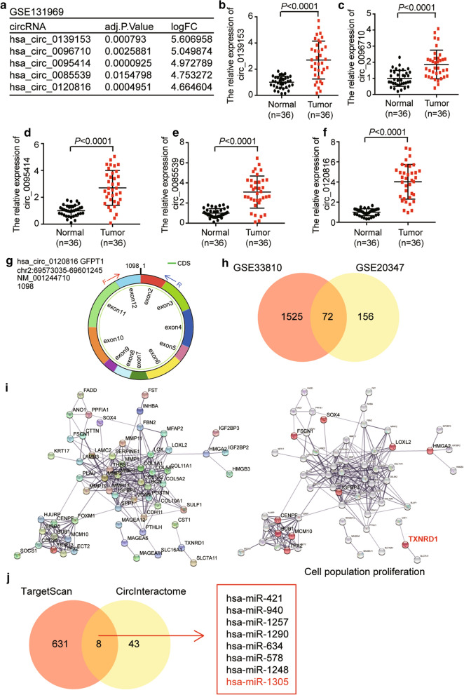
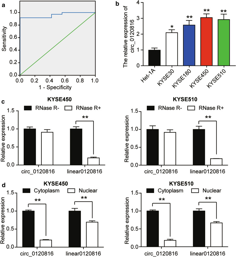
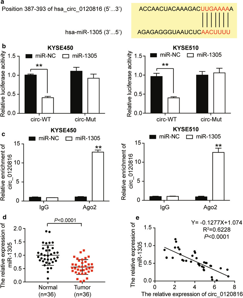
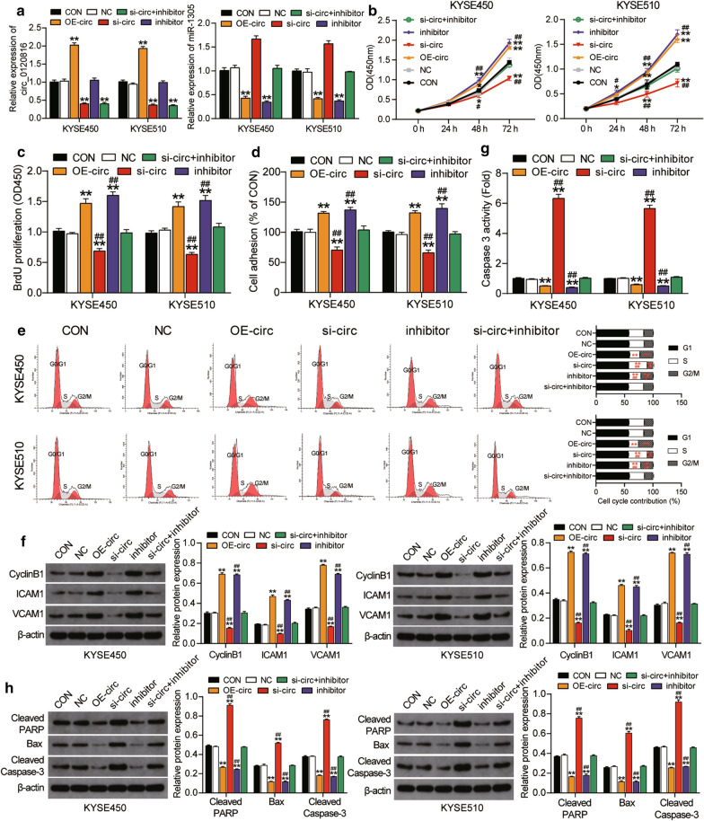
Methods: Bioinformatics analysis was first performed to screen dysregulated circRNAs and differentially expressed genes in ESCC. The ESCC tissue samples and adjacent normal tissue samples utilized in this study were obtained from 36 ESCC patients. All the samples were subjected to qRT-PCR analysis to identify the expression of TXNRD1, circRNAs, and miR-1305. Luciferase reporter assay, RNA immunoprecipitation assay and RNA pull-down assay were later conducted to verify the existing relationship among circ0120816, miR-1305 and TXNRD1. CCK-8, BrdU, cell adhesion, cell cycle, western blot and caspase 3 activity assays were also employed to evaluate the regulation of these three biological molecules in ESCC carcinogenesis. To evaluate the effect of circ0120816 on ESCC tumor growth and metastasis, the xenograft mice model was constructed.

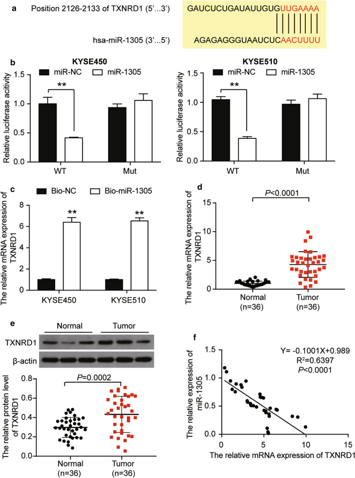
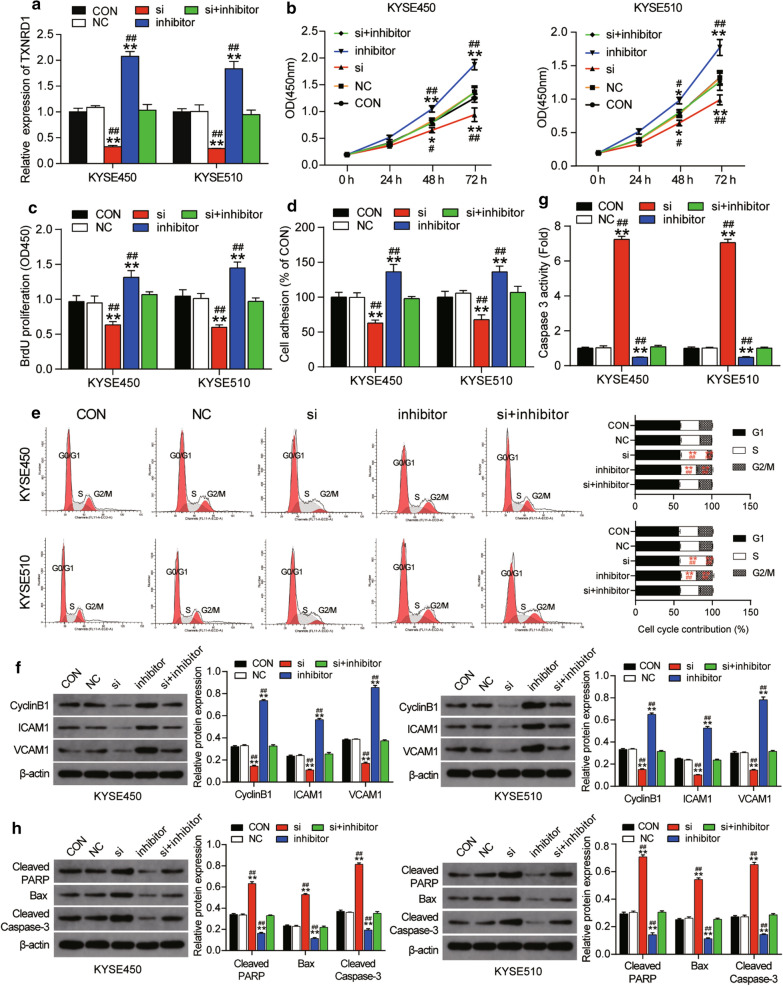
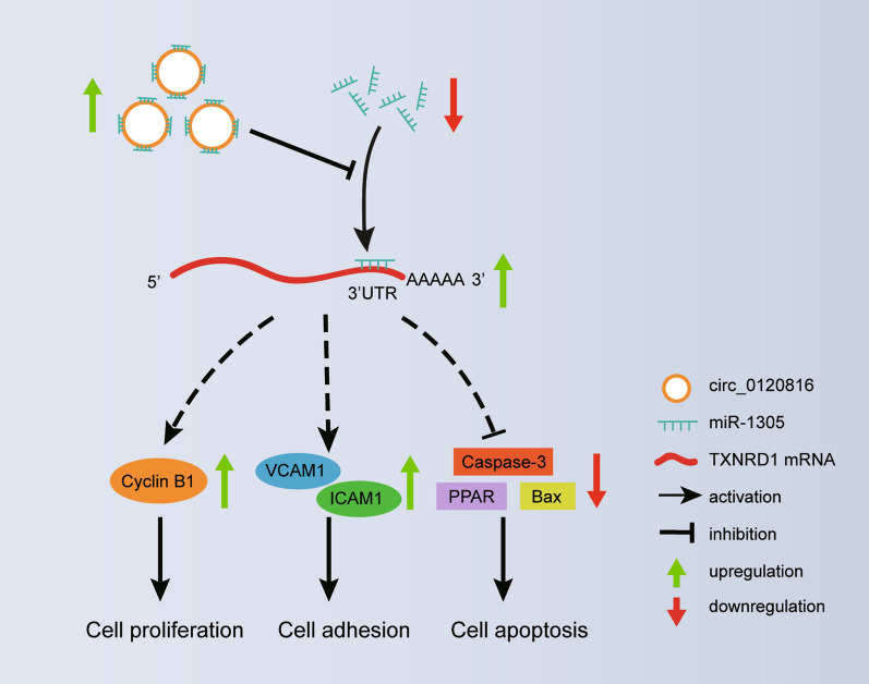
Results: Experimental investigations revealed that circ0120816 was the highest upregulated circRNA in ESCC tissues and that this non-coding RNA acted as a miR-1305 sponge in enhancing cell viability, cell proliferation, and cell adhesion as well as repressing cell apoptosis in ESCC cell lines. Moreover, miR-1305 was observed to exert a tumor-suppressive effect in ESCC cells by directly targeting and repressing TXNRD1. It was also noticed that TXNRD1 could regulate cyclin, cell adhesion molecule, and apoptosis-related proteins. Furthermore, silencing circ0120816 was found to repress ESCC tumor growth and metastasis in vivo.

Conclusions: This research confirmed that circ0120816 played an active role in promoting ESCC development by targeting miR-1305 and upregulating oncogene TXNRD1.

Keywords: Esophageal squamous cell carcinoma; TXNRD1; circ0120816; miR-1305.


Conflict of interest statement

The authors declare that no conflict of interests exist in this research project.

Figures

Fig. 1
Fig. 1
The identification of circ0120816/miR-1305/TXNRD1 axis in ESCC. a Microarray analysis revealed the top 5 upregulated circRNAs in circRNA microarray GSE131969. bf RT-qPCR analysis showed the expression of the top 5 upregulated circRNAs in ESCC clinical samples. g The structure of circ0120816. h A total of 72 upregulated DEGs were overlapped in datasets of GSE33810 and GSE20347 using Venny 2.1.0 analysis. i TXNRD1 was the key gene involved in cell population proliferation. j Eight miRNAs were identified to be overlapped using TargetScan and circInteractome analyses. TargetScan was utilized to predict miRNAs binding to TXNRD1. CircInteractome was employed to predict miRNAs binding to circ0120816
Fig. 2
Fig. 2
Circ0120816 was upregulated in ESCC cells. a The receiver operating characteristic (ROC) curve showed the diagnostic value of circ0120816 for ESCC. The ROC curve was determined based on the circ0120816 expression in ESCC tissues and adjacent normal tissues. b RT-qPCR analysis revealed the expression of circ0120816 in ESCC cell lines (KYSE30, KYSE180, KYSE450, KYSE510) and normal cells (Het-1A). *P < 0.05, **P < 0.001, compared with Het-1A cells. c RNase R degradation assay confirmed the stability of circ0120816. **P < 0.001, the comparison between RNase R- and RNase R + . d The distribution of circ0120816 and linear 0120816 in ESCC cells was determined by circRNA subcellular localization assay. **P < 0.001, the comparison between the cytoplasm and the nucleus. Each cellular experiment was independently repeated three times, and the data collected were displayed in the format of mean ± standard deviation (SD)
Fig. 3
Fig. 3
Circ0120816 acted as a sponge for miR-1305. a The binding sequence between circ0120816 and miR-1305 was predicted by TargetScan 7.0. b Luciferase reporter assay was used to confirm the relationship between circ0120816 and miR-1305. **P < 0.001, compared with miR-NC. miR-NC: miR-1305 negative control, circ-WT: circ0120816 wild type, circ-Mut: circ0120816 mutant type. c RIP analysis showed that circ0120816 was abundantly pulled down by antiAgo2 antibodies when transfected with miR-1305 mimics in ESCC cells compared with the miR-1305 NC and IgG group. **P < 0.001, compared with miR-NC. miR-NC: miR-1305 negative control, IgG: negative control, Ago2: anti-Argonaute2 antibody. d RT-qPCR analysis showed that the expression of miR-1305 was decreased in ESCC tissues. Normal: adjacent healthy tissues, Tumor: ESCC tissues. e Pearson’s correlation analysis showed that miR-1305 was negatively associated with circ0120816. Each cellular experiment was independently repeated three times, and the data collected were illustrated in the format of mean ± standard deviation (SD)
Fig. 4
Fig. 4
Circ0120816 facilitated ESCC progression by sponging miR-1305. a RT-qPCR analysis of the expression of circ0120816 and miR-1305 in ESCC cell lines after transfecting with circ0120816 overexpression plasmid, circ0120816 siRNA or miR-1305 inhibitor. b CCK-8 assay was used to determine the viability of ESCC cell lines after transfection. c BrdU assay was conducted to evaluate the proliferation of ESCC cell lines after transfection. d Cell adhesion assay was employed to determine the cell adhesion ability of ESCC cell lines after transfection. e Cell cycle assay was applied to evaluate the cell cycle in ESCC cell lines after transfecting with a flow cytometry. f The expression of cyclinB1, ICAM1 and VCAM1 was detected in ESCC cell lines after transfection by western blot assay. g Caspase 3 activity assay indicates the cell apoptosis of ESCC cell lines after transfection. h The expression of Cleaved PARP, Bax and Cleaved Caspase-3 was detected in ESCC cell lines after transfecting with western blot assay. *P < 0.05, **P < 0.001, compared with the blank control group. #P < 0.05, ##P < 0.001, compared with the co-transfection group of si-circ0120816 plus miR-1305 inhibitor. CON: blank control, NC: negative control, OE-circ: circ0120816 overexpression plasmid, si-circ: circ0120816 siRNA, inhibitor: miR-1305 inhibitor, si-circ + inhibitor: circ0120816 siRNA plus miR-1305 inhibitor. Each experiment was independently repeated three times, and the data collected were displayed in the format of mean ± standard deviation (SD)
Fig. 5
Fig. 5
Silencing circ0120816 could inhibit the tumor growth and lung metastasis of ESCC in vivo. a Representative live imaging of xenograft mice with transplantation of KYSE450 cells lines pre-transfected with circ0120816 shRNA (Sh-circ) or negative control (NC) at week 1 and week 4. b Statistical analysis of the live imaging results. c RT-qPCR analysis of circ0120816 expression in the tumor tissues of xenograft mice. d RT-qPCR analysis of miR-1305 expression in the tumor tissues of xenograft mice. e Pathological H&E staining of tumor tissues collected from the Sh-circ and NC group mice. f Pathological H&E staining of lung tissues collected from the Sh-circ and NC group mice. **p < 0.001 compared with the NC group
Fig. 6
Fig. 6
TXNRD1 was a target gene of miR-1305. a The binding sequence between TXNRD1 3′UTR and miR-1305 was predicted by TargetScan 7.0. b Luciferase reporter assay was used to observe the relationship between miR-1305 and TXNRD1. **P < 0.001, compared with miR-NC. miR-NC: miR-1305 negative control, WT: TXNRD1 wild type, Mut: TXNRD1 mutant type. c RNA pull-down assay was used to determine the relationship between miR-1305 and TXNRD1. **P < 0.001, compared with Bio-NC. Bio-NC: Bio-miR-1305 negative control. Bio-miR-1305: miR-1305 mimic-biotin. d RT-qPCR analysis showed that the expression of TXNRD1 was increased in ESCC tissues. e Pearson’s correlation analysis revealed that MiR-1305 had a negative relationship with TXNRD1. Each cellular experiment was independently repeated three times, and the data collected were displayed in the format of mean ± standard deviation (SD)
Fig. 7
Fig. 7
MiR-1305 suppressed ESCC progression by targeting TXNRD1. a RT-qPCR analysis of the expression of TXNRD1 after transfecting with TXNRD1 siRNA and/or miR-1305 inhibitor. b CCK-8 assay was performed to evaluate the cell viability of ESCC cells after transfection. c BrdU assay was conducted to evaluate the proliferation of ESCC cell lines after transfection. d Cell adhesion assay was used to assess the adhesion ability of ESCC cell lines after transfection. e Cell cycle assay was applied to evaluate the cell cycle in ESCC cell lines after transfecting with a flow cytometry system. f The expression of cyclinB1, ICAM1 and VCAM1 was detected in ESCC cell lines after transfecting with western blot assay. g Caspase 3 activity assay indicates the cell apoptosis of ESCC cell lines after transfection. h The expression of Cleaved PARP, Bax and Cleaved Caspase-3 was detected in ESCC cell lines after transfecting with western blot assay. *P < 0.05, **P < 0.001, compared with the blank control group. #P < 0.05, ##P < 0.001, compared with the co-transfection group of si-TXNRD1 plus miR-1305 inhibitor. CON: blank control, NC: negative control, inhibitor: miR-1305 inhibitor, si: TXNRD1 siRNA, si + inhibitor: TXNRD1 siRNA plus miR-1305 inhibitor. Each experiment was independently repeated three times, and the data collected were displayed in the format of mean ± standard deviation (SD)
Fig. 8
Fig. 8
The schematic diagram of circ0120816/miR-1305/TXBRD1 axis working in ESCC tumorigenesis

Similar articles

Cited by

References

    1. Bray F, Ferlay J, Soerjomataram I, Siegel RL, Torre LA, Jemal A. Global cancer statistics 2018: GLOBOCAN estimates of incidence and mortality worldwide for 36 cancers in 185 countries. CA Cancer J Clin. 2018;68(6):394–424. - PubMed
    1. Uhlenhopp DJ, Then EO, Sunkara T, Gaduputi V. Epidemiology of esophageal cancer: update in global trends, etiology and risk factors. Clin J Gastroenterol. 2020 doi: 10.1007/s12328-020-01237-x. - DOI - PubMed
    1. Wei WQ, Chen ZF, He YT, Feng H, Hou J, Lin DM, Li XQ, Guo CL, Li SS, Wang GQ, et al. Long-term follow-up of a community assignment, one-time endoscopic screening study of esophageal cancer in China. J Clin Oncol. 2015;33(17):1951–1957. doi: 10.1200/JCO.2014.58.0423. - DOI - PMC - PubMed
    1. Saxena R, Klochkova A, Murray MG, Kabir MF, Samad S, Beccari T, Gang J, Patel K, Hamilton KE, Whelan KA. Roles for autophagy in esophageal carcinogenesis: implications for improving patient outcomes. Cancers (Basel) 2019;11(11):1697. doi: 10.3390/cancers11111697. - DOI - PMC - PubMed
    1. Riley RS, June CH, Langer R, Mitchell MJ. Delivery technologies for cancer immunotherapy. Nat Rev Drug Discovery. 2019;18(3):175–196. doi: 10.1038/s41573-018-0006-z. - DOI - PMC - PubMed