. 2020 Nov 27;20(1):571.
doi: 10.1186/s12935-020-01665-2.

Long non-coding RNA NORAD contributes to the proliferation, invasion and EMT progression of prostate cancer via the miR-30a-5p/RAB11A/WNT/β-catenin pathway

Affiliations

Long non-coding RNA NORAD contributes to the proliferation, invasion and EMT progression of prostate cancer via the miR-30a-5p/RAB11A/WNT/β-catenin pathway

Yunxia Zhang et al. Cancer Cell Int. .

Abstract

Background: Prostate cancer (PC) is common male cancer with high mortality worldwide. Emerging evidence demonstrated that long noncoding RNAs (lncRNAs) play critical roles in various type of cancers including PC by serving as competing endogenous RNAs (ceRNAs) to modulate microRNAs (miRNAs). LncRNA activated by DNA damage (NORAD) was found to be upregulated in PC cells, while the detailed function and regulatory mechanism of NORAD in PC progression remains largely unclear.

Methods: Expression of NORAD in PC tissues and cell lines were detected by real-time quantitative PCR (qRT-PCR). NORAD was respectively overexpressed and knocked down by transfection with pcDNA-NORAD and NORAD siRNA into PC-3 and LNCap cells. Cell proliferation, invasion and apoptosis were determined by using CCK-8, Transwell and Flow cytometry assays, respectively. The target correlations between miR-30-5p and NORAD or RAB11A were confirmed by using dual luciferase reporter assay. Moreover, expression levels of RAB11A, the epithelial-mesenchymal transition (EMT) marker proteins and the Wnt pathway related proteins were measured by Western blotting. Tumor xenograft assay was used to study the effect of NORAD on tumor growth in vivo.

Results: NORAD was upregulated in PC tissues and cells. Overexpression of NORAD promoted cell proliferation, invasion, EMT, and inhibited cell apoptosis; while knockdown of NORAD had the opposite effect. NORAD was found to be functioned as a ceRNA to bind and downregulated miR-30a-5p that was downregulated in PC tumor tissues. Rescue experiments revealed that miR-30a-5p could weaken the NORAD-mediated promoting effects on cell proliferation, invasion and EMT. Furthermore, RAB11A that belongs to a member of RAS oncogene family was verified as a target of miR-30a-5p, and reintroduction of RAB11A attenuated the effects of miR-30a-5p overexpression on cell proliferation, invasion, EMT and apoptosis of PC cells. More importantly, silencing RAB11A partially reversed the promoting effects of NORAD overexpression on cell proliferation, invasion and EMT of PC cells via the WNT/β-catenin pathway. Lastly, tumorigenicity assay in vivo demonstrated that NORAD increased tumor volume and weight via miR-30a-5p /RAB11A pathway.

Conclusion: Our results indicated a significant role of NORAD in mechanisms associated with PC progression. NORAD promoted cell proliferation, invasion and EMT via the miR-30a-5p/RAB11A/WNT/β-catenin pathway, thus inducing PC tumor growth.

Keywords: Apoptosis; Epithelial–mesenchymal transition; Proliferation; Prostate cancer; RAB11A; Wnt/β-catenin; lncRNA NORAD; miR-30a-5p.


Conflict of interest statement

The authors declare that they have no competing interests.

Figures

Fig. 1
Fig. 1
The expression level of LncRNA NORAD in PC tumor tissues and cell lines. a The relative expression levels of NORAD in PC tissues (Tumor tissues, n = 45) and adjacent normal tissues (Normal tissues, n = 45) determined by qRT-PCR. b Relative expression of NORAD was detected by qRT-PCR in PC cell lines (PC-3, LNCap, 22RV1, DU-145). The normal human prostate epithelial cell line (RWPE-1) served as a control. n = 3. The data were presented as the mean ± standard error of mean (SEM) and Student’s t-test was used for the comparison between 2 groups. *P < 0.05
Fig. 2
Fig. 2
Effects of NORAD overexpression on cell proliferation, invasion, apoptosis, and EMT process of PC-3 cells. PC-3 cells were transfected with 0.5 or 2 μg/mL pcDNA-NORAD expression vector, or negative control Vector (0.5 μg/mL). a Expression of NORAD was then determined by qRT-PCR after 48 h transfection. b Cell proliferation of PC-3 cells after infection for 24, 48, 72 and 96 h were detected by CCK-8 assay. c, d Transwell and Flow cytometry was performed to determine cell invasion and apoptosis in PC-3 cells after infection for 48 h, respectively. e The expression levels of N-cadherin, E-cadherin, Vimentin and Snail in PC-3 cells after infection with pcDNA-NORAD for 48 h were detected by Western blotting. The data were presented as the mean ± standard error of mean (SEM), n = 3. Student’s t-test was used for the comparison between 2 groups, and one-way analysis of variance (ANOVA) was used for the comparison among more than 2 groups in this study. *P < 0.05
Fig. 3
Fig. 3
Effects of silencing NORAD on cell proliferation, invasion, apoptosis, and EMT process of PC-3 cells. PC-3 cells were transfected with 10 nM or 30 nM NORAD small interfering siRNA (NORAD siRNA), or negative control (10 nM). a Expression of NORAD was then determined by qRT-PCR after 48 h transfection. b Cell proliferation of PC-3 cells after infection for 24, 48, 72 and 96 h were detected by CCK-8 assay. c, d Transwell and Flow cytometry was performed to determine cell invasion and apoptosis in PC-3 cells after infection for 48 h, respectively. e The expression levels of N-cadherin, E-cadherin, Vimentin and Snail in PC-3 cells after transfection with NORAD siRNAs for 48 h were detected by Western blotting. The data were presented as the mean ± standard error of mean (SEM), n = 3. Student’s t-test was used for the comparison between 2 groups, and one-way analysis of variance (ANOVA) was used for the comparison among more than 2 groups in this study. *P < 0.05
Fig. 4
Fig. 4
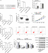
Reintroduction of miR-30a-5p weakens the effect of NORAD overexpression on cell proliferation, invasion, apoptosis and EMT process of PC-3 cells. a The potential binding sequences between NORAD and miR-30a-5p predicted by starBase. b qRT-PCR was conducted to determine the expression levels of miR-30a-5p in PC-3 cells transfected with 100 nM miR-30a-5p mimic, 100 nM miR-30a-5p inhibitor, or 100 nM their negative controls for 48 h. c Dual luciferase reporter assay was performed to determine the luciferase activity of HEK 293 T cells transfected with miR-30a-5p mimic and luciferase reporter vectors containing WT- or MUT- NORAD segment. d The expression level of miR-30a-5p was detected by qRT-PCR in PC-3 cell after being transfected with 2 μg/mL pcDNA-NORAD, 30 nM NORAD siRNA, or their negative controls for 48 h. e qRT-PCR was performed to determine the expression levels of miR-30a-5p in PC tissues (Tumor tissues, n = 45) and adjacent normal tissues (Normal tissues, n = 45) determined by qRT-PCR. Then, PC-3 cells were transfected with 2 μg/mL pcDNA-NORAD alone, or together with 100 nM miR-30a-5p mimic. f Cell proliferation of PC-3 cells after infection for 24, 48, 72 and 96 h were detected by CCK-8 assay. g, h After 48 h transfection, cell apoptosis and invasion was determined by Flow cytometry and Transwell, respectively. i The expression levels of N-cadherin, E-cadherin, Vimentin and Snail in PC-3 cells after infection of 48 h were detected by Western blotting. The data were presented as the mean ± standard error of mean (SEM). Student’s t-test was used for the comparison between 2 groups in this study. *P < 0.05
Fig. 5
Fig. 5
Effects of miR-30a-5p on cell proliferation, invasion, apoptosis and EMT in PC-3 cells by targeting RAB11A. a Diagram of the predicted miR-30a-5p binding sites in the 3′-UTR of RAB11A mRNA by TargetScan. b Dual luciferase reporter assay was performed to detect whether miR-30a-5p directly bound to the predicted binding sites in 3′-UTR of RAB11A mRNA. c The relative expression levels of RAB11A mRNA were determined by qRT-PCR in PC tissues (Tumor tissues, n = 45) and adjacent normal tissues (Normal tissues, n = 45) determined by qRT-PCR. d, e qRT-PCR and Western blotting assays were conducted to determine the mRNA and protein expression levels of RAB11A in PC-3 cells transfected with 100 nM miR-30a-5p mimic, 100 nM miR-30a-5p inhibitor, or 100 nM their negative controls for 48 h. f, g After infection PC-3 cells with RAB11A overexpression vector (1.5 μg/mL) for 48 h, the expression of RAB11A was determined by qRT-PCR and Western blotting at mRNA and protein levels. Then, PC-3 cells were transfected with 100 nM miR-30a-5p mimic alone, or together with 1.5 μg/mL RAB11A overexpression vector. h CCK-8 was used to detect the cell proliferation of PC-3 cells after transfectio for 48 h. i, j Flow cytometry and Transwell was performed to determined apoptosis and invasion in PC-3 cells after 48 h transfection. k The expression levels of N-cadherin, E-cadherin, Vimentin and Snail were detected by Western blotting in transfected PC-3 cells. The data were presented as the mean ± standard error of mean (SEM). Student’s t test was used for the comparison between 2 groups in this study. *P < 0.05
Fig. 6
Fig. 6
NORAD promotes cell proliferation, invasion and EMT, suppresses cell apoptosis via miR-30a-5p/RAB11A/WNT/β-catenin pathway in PC-3 cells. PC-3 cells were transfected with 50 nM RAB11A siRNA for 48 h. a, b Interference efficiencies of RAB11A siRNA were determined by qRT-PCR and Western blotting, respectively. Subsequently, PC-3 cells were transfected with 2 μg/mL pcDNA-NORAD alone, or together with 50 nM RAB11A siRNA. c After 48 h transfection, CCK-8 was used to detect the cell proliferation. d, e Cell invasion and apoptosis was determined by Transwell and Flow cytometry in PC-3 cells after 48 h transfection with NORAD alone or together with RAB11A siRNA. f, g Western blotting was used to determine the expression levels of related proteins of EMT (N-cadherin, E-cadherin, Vimentin and Snail) and WNT pathway (β-catenin, Cyclin D and c-Myc) in PC-3 cells after 48 h transfection. The data were presented as the mean ± standard error of mean (SEM), n = 3. Student’s t-test was used for the comparison between 2 groups in this study. *P < 0.05
Fig. 7
Fig. 7
Downregulation of NORAD suppresses tumor growth in vivo. PC-3 cells were transfected with NORAD siRNA alone, or together with miR-30a-5p inhibitor for 48 h. Then, the transfected PC-3 cells were subcutaneously injected into the right flanks of nude mice. a The tumor volume was measured once a week for four weeks and calculated by the formula of 0.5 × Length × Width2. b Tumor-bearing mice were killed after four weeks’ cultivation, and the weight of the tumor was weighed in each group. c, d The expression levels of NORAD and miR-30a-5p were determined by qRT-PCR in tumor tissues harvested from the nude mice in each group. e, f Western blotting was used to detect the expression of RAB11A protein and the related proteins of WNT pathway (β-catenin, Cyclin D and c-Myc) in tumor tissues harvested from the nude mice in each group. The total number of mice is eighty (n = 5/group). The data were presented as the mean ± standard error of mean (SEM). Student’s t-test was used for the comparison between 2 groups in this study. *P < 0.05

Similar articles

Cited by

References

    1. Davidsson S, Andren O, Ohlson AL, Carlsson J, Andersson SO, Giunchi F, Rider JR, Fiorentino M. FOXP3(+) regulatory T cells in normal prostate tissue, postatrophic hyperplasia, prostatic intraepithelial neoplasia, and tumor histological lesions in men with and without prostate cancer. Prostate. 2018;78(1):40–47. doi: 10.1002/pros.23442. - DOI - PMC - PubMed
    1. Shi X, Zhang W, Nian X, Lu X, Li Y, Liu F, Wang F, He B, Zhao L, Zhu Y, Ren S, Sun Y. The previously uncharacterized lncRNA APP promotes prostate cancer progression by acting as a competing endogenous RNA. Int J Cancer. 2020;146(2):475–486. doi: 10.1002/ijc.32422. - DOI - PubMed
    1. Zhang J, Wang L, You X, Xian T, Wu J, Pang J. Nanoparticle therapy for prostate cancer: overview and perspectives. Curr Top Med Chem. 2019;19(1):57–73. doi: 10.2174/1568026619666190125145836. - DOI - PubMed
    1. Song C, Chen H. Predictive significance of TMRPSS2-ERG fusion in prostate cancer: a meta-analysis. Cancer Cell Int. 2018;18:177. doi: 10.1186/s12935-018-0672-2. - DOI - PMC - PubMed
    1. Wang J, Yang Y, Li K. Long noncoding RNA DANCR aggravates retinoblastoma through miR-34c and miR-613 by targeting MMP-9. J Cell Physiol. 2018;233(10):6986–6995. doi: 10.1002/jcp.26621. - DOI - PubMed

LinkOut - more resources