Review
. 2020 Nov 20;17(1):38.
doi: 10.1186/s12979-020-00208-7.

Autoimmune hemolytic anemia: current knowledge and perspectives

Affiliations
Review

Autoimmune hemolytic anemia: current knowledge and perspectives

Sylwia Sulimiera Michalak et al. Immun Ageing. .

Abstract

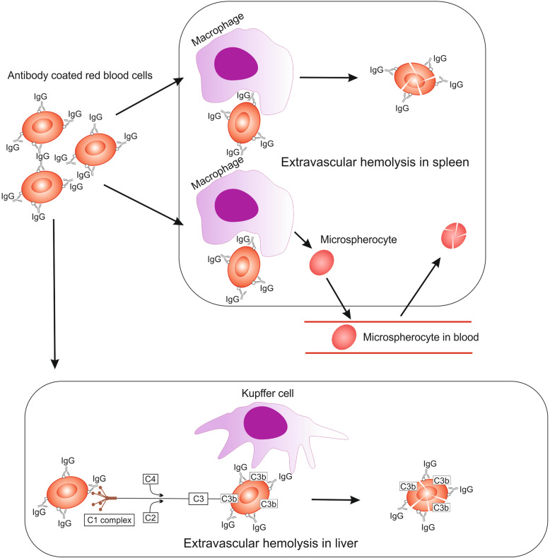
Autoimmune hemolytic anemia (AIHA) is an acquired, heterogeneous group of diseases which includes warm AIHA, cold agglutinin disease (CAD), mixed AIHA, paroxysmal cold hemoglobinuria and atypical AIHA. Currently CAD is defined as a chronic, clonal lymphoproliferative disorder, while the presence of cold agglutinins underlying other diseases is known as cold agglutinin syndrome. AIHA is mediated by autoantibodies directed against red blood cells (RBCs) causing premature erythrocyte destruction. The pathogenesis of AIHA is complex and still not fully understood. Recent studies indicate the involvement of T and B cell dysregulation, reduced CD4+ and CD25+ Tregs, increased clonal expansions of CD8 + T cells, imbalance of Th17/Tregs and Tfh/Tfr, and impaired lymphocyte apoptosis. Changes in some RBC membrane structures, under the influence of mechanical stimuli or oxidative stress, may promote autohemolysis. The clinical presentation and treatment of AIHA are influenced by many factors, including the type of AIHA, degree of hemolysis, underlying diseases, presence of concomitant comorbidities, bone marrow compensatory abilities and the presence of fibrosis and dyserthropoiesis. The main treatment for AIHA is based on the inhibition of autoantibody production by mono- or combination therapy using GKS and/or rituximab and, rarely, immunosuppressive drugs or immunomodulators. Reduction of erythrocyte destruction via splenectomy is currently the third line of treatment for warm AIHA. Supportive treatment including vitamin supplementation, recombinant erythropoietin, thrombosis prophylaxis and the prevention and treatment of infections is essential. New groups of drugs that inhibit immune responses at various levels are being developed intensively, including inhibition of antibody-mediated RBCs phagocytosis, inhibition of B cell and plasma cell frequency and activity, inhibition of IgG recycling, immunomodulation of T lymphocytes function, and complement cascade inhibition. Recent studies have brought about changes in classification and progress in understanding the pathogenesis and treatment of AIHA, although there are still many issues to be resolved, particularly concerning the impact of age-associated changes to immunity.

Keywords: Autoimmune hemolytic anemia; Cold agglutinin disease; Microvesicles; Pathogenesis; Shear stress; Treatment.


Conflict of interest statement

The authors declare that they have no conflict of interest.

Figures

Fig. 1
Fig. 1
A schematic visualization of RBCs exposed to shear stress
Fig. 2
Fig. 2
A schematic visualization of erythrocyte senescence and microvesicle generation
Fig. 3
Fig. 3
Hemolysis in the course of warm AIHA: the main pathological mechanisms and sites of hemolysis, adapted and modified from Berentsen and Sundic [72]. Antibody-coated RBCs are ingested and degraded by macrophages or destroyed by antibody dependent cell cytotoxicity in the spleen. Some of the erythrocytes are only partially phagocytosed and form microspherocytes which return to the circulation. Microspherocytes have rigid cellular membrane and lack flexibility which causes their sequestration in the spleen. Weak activation of complement by Ig G antibodies leads to extravascular hemolysis in the liver
Fig. 4
Fig. 4
Hemolysis in CAD, adapted and modified from Berentsen et al. [72, 74]. Activation of the complement cascade caused by the reaction of cold agglutinins with RBCs leads to intravascular and extravascular hemolysis. Extravascular hemolysis is associated with complement activation and destruction of RBCs by the mononuclear phagocytic system, mainly in the liver. Intravascular hemolysis is a result of the formation of membrane attack complex (MAC), composed of C5b, C6, C7, C8, and C9
Fig. 5
Fig. 5
New, potential drugs targeting mainly immune cells, adapted and modified from Michalak [108]. BTK - Bruton’s tyrosine kinase, CD - cluster of differentiation - cell surface antigen, FcRn - neonatal crystallizable fragment of the receptor, IL 2 - interleukin 2, IL 2R - interleukin 2 receptor, mTOR - mammalian target of rapamycin kinase, PI3K - hosphatidylinositol 3-kinase, PI3K/AKT - intracellular signaling pathway, SYK - spleen tyrosine kinase, SYNT001 - monoclonal antibody (Orilanolimab)

References

    1. Goronzy JJ, Li G, Yang Z, Weyand CM. The janus head of T cell aging - autoimmunity and immunodeficiency. Front Immunol. 2013;4:131. doi: 10.3389/fimmu.2013.00131. - DOI - PMC - PubMed
    1. Barcellini W, Fattizzo B, Zaninoni A. Current and emerging treatment options for autoimmune hemolytic anemia. Expert Rev Clin Immunol. 2018;14(10):857–872. doi: 10.1080/1744666X.2018.1521722. - DOI - PubMed
    1. Jäger U, Barcellini W, Broome CM, Gertz MA, Hill A, Hill QA, et al. Diagnosis and treatment of autoimmune hemolytic anemia in adults: Recommendations from the First International Consensus Meeting. Blood Rev. 2020;41:100648. doi: 10.1016/j.blre.2019.100648. - DOI - PubMed
    1. Hill QA, Hill A, Berentsen S. Defining autoimmune hemolytic anemia: a systematic review of the terminology used for diagnosis and treatment. Blood Adv. 2019;3(12):1897–1906. doi: 10.1182/bloodadvances.2019000036. - DOI - PMC - PubMed
    1. Lazarian G, Quinquenel A, Bellal M, Siavellis J, Jacquy C, Re D, et al. Autoimmune haemolytic anaemia associated with COVID-19 infection. Br J Haematol. 2020;190(1):29–31. doi: 10.1111/bjh.16794. - DOI - PMC - PubMed