. 2020 Nov 23;10(1):134.
doi: 10.1186/s13578-020-00497-x.

CXCR7: a β-arrestin-biased receptor that potentiates cell migration and recruits β-arrestin2 exclusively through Gβγ subunits and GRK2

Affiliations

CXCR7: a β-arrestin-biased receptor that potentiates cell migration and recruits β-arrestin2 exclusively through Gβγ subunits and GRK2

Huong Thi Nguyen et al. Cell Biosci. .

Abstract

Background: Some chemokine receptors referred to as atypical chemokine receptors (ACKRs) are thought to non-signaling decoys because of their inability to activate typical G-protein signaling pathways. CXCR7, also known as ACKR3, binds to only two chemokines, SDF-1α and I-TAC, and recruits β-arrestins. SDF-1α also binds to its own conventional receptor, CXCR4, involving in homeostatic modulation such as development and immune surveillance as well as pathological conditions such as inflammation, ischemia, and cancers. Recently, CXCR7 is suggested as a key therapeutic target together with CXCR4 in such conditions. However, the molecular mechanisms underlying cellular responses and functional relation with CXCR7 and CXCR4 have not been elucidated, despite massive studies. Therefore, we aimed to reveal the molecular networks of CXCR7 and CXCR4 and compare their effects on cell migration.

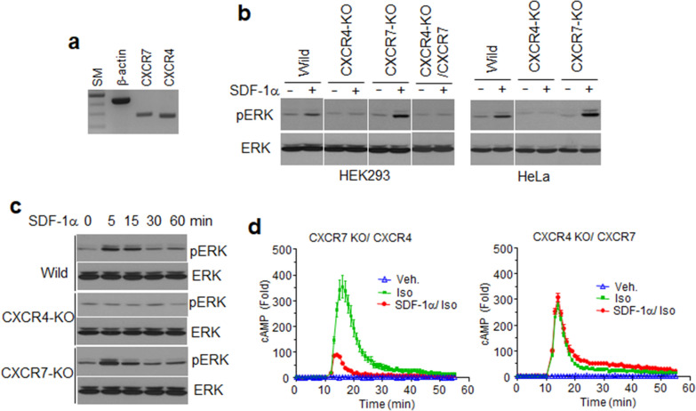
Methods: Base on structural complementation assay using NanoBiT technology, we characterized the distinct mechanisms underlying β-arrestin2 recruitment by both CXCR4 and CXCR7. Crosslinking and immunoprecipitation were conducted to analyze complex formation of the receptors. Gene deletion using CRISPR and reconstitution of the receptors were applied to analysis of ligand-dependent ERK phosphorylation and cell migration. All experiments were performed in triplicate and repeated more than three times. Unpaired Student's t-tests or ANOVA using PRISM5 software were employed for statistical analyses.

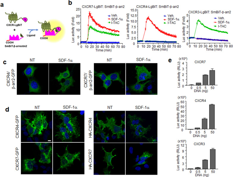
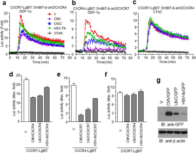
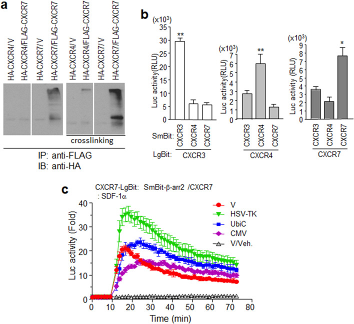
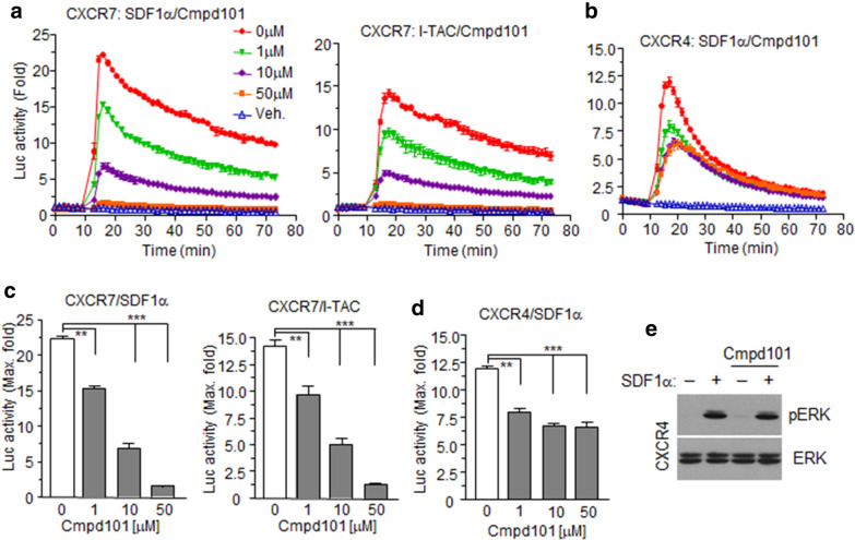
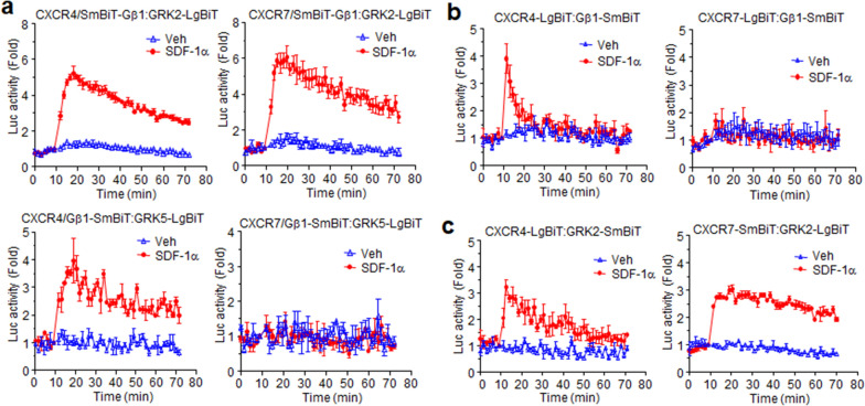
Results: Ligand binding to CXCR7 does not result in activation of typical signaling pathways via Gα subunits but activation of GRK2 via βγ subunits and receptor phosphorylation with subsequent β-arrestin2 recruitment. In contrast, CXCR4 induced Gαi activation and recruited β-arrestin2 through C-terminal phosphorylation by both GRK2 and GRK5. SDF-1α-stimulated ERK phosphorylation was facilitated by CXCR4, but not CXCR7. Heterodimerization of CXCR4 and CXCR7 was not confirmed in this study, while homodimerization of them was verified by crosslinking experiment and NanoBiT assay. Regarding chemotaxis, SDF-1α-stimulated cell migration was mediated by both CXCR4 and CXCR7.

Conclusion: This study demonstrates that SDF-1α-stimulated CXCR7 mediates β-arrestin2 recruitment via different molecular networking from that of CXCR4. CXCR7 may be neither a simple scavenger nor auxiliary receptor but plays an essential role in cell migration through cooperation with CXCR4.

Keywords: Biased GPCR; CXCR4; CXCR7; Chemotaxis; SDF-1α; Structural complementation assay.


Conflict of interest statement

The authors declare that they have no competing interests.

Figures

Fig. 1
Fig. 1
SDF-1α induced β-arrestin2 recruitment to CXCR4 and CXCR7. a Schematic representation of the structural complementation assays for the interaction of β-arrestin2 with chemokine receptors b HEK293 cells, transiently transfected with constructs for receptor-LgBiT and SmBiT-β-arrestin2, were treated with the indicated chemokines (I-TAC/SDF-1α) or not (Veh.), and then assessed for real-time luminescence. c CXCR4 and CXCR7 mediate intracellular clustering of β-arrestin2 by SDF-1α. Cells transfected with β-arrestin2-GFP and chemokine receptor constructs were treated with SDF-1α for 30 min. GFP fluorescence was then assessed using confocal microscopy. d Translocation of CXCR4 and CXCR7 to the cytosol by SDF-1α. Cells expressing CXCR4-GFP or CXCR7-GFP were treated with SDF-1α and fixed. Confocal images show the subcellular localization of GFP signals (left panels). Cells expressing HA-tagged receptors were labeled with anti-HA antibodies and treated with SDF-1α for 30 min (NT: no treatment). Fixed cells were permeabilized and stained with FITC-conjugated goat anti-mouse IgG. The images show the subcellular localization of the HA-tagged receptors (right panel). e Cell surface expression of the receptors. Transfected cells with constructs for HiBiT-tagged receptors were incubated with extracellular HiBiT detection reagent, and luminescence was measured. The results are an average of three independent experiments. Values are presented as the mean ± SD
Fig. 2
Fig. 2
CXCR7 and CXCR4 compete with each other for SDF-1α ligand specificity. ac NanoBiT receptor/β-arrestin2 constructs were co-expressed with other receptors governed by different promoters as described for each graph. df Graphs of maximum luminescence activities in each experiment from (ac). g Transcriptional activities of each promoter. HEK293 cells expressing GFP governed by the CMV (CMV/GFP), Ubiquitin C (UbiC/GFP), or herpes simplex virus-thymidine kinase (HSV-tk/GFP) promoter were lysed and used for western blotting with anti-GFP antibodies. Results are the average of three independent experiments. Values are presented as the mean ± SD
Fig. 3
Fig. 3
Homodimerization of CXCR7 on the membrane surface. a HEK293 cells expressing epitope-tagged forms of CXCR4 or CXCR7 were used for immunoprecipitation with anti-FLAG agarose and subsequent western blot analysis with anti-HA antibodies. Cross-linking experiments were performed as described in Materials and Methods. b Luminescence produced by receptor dimerization. NanoBiT constructs for each receptor were expressed in HEK293 cells. c NanoBiT constructs of CXCR7 and β-arrestin2 were co-expressed with different promoter constructs of CXCR7 (HSV-TK, UbiC, CMV) or empty vector (V) in HEK293 cells. Cells were treated with SDF-1α or not (V/Veh), and then luminescence was measured. Results are the average of three independent experiments. Values are presented as the mean ± SD. *p < 0.05, **p < 0.001
Fig. 4
Fig. 4
Characterization of receptor-mediated chemokine signaling events. a cAMP assay. HEK293 cells transfected with receptor gene plasmids or an empty vector (V) and pGlo22F plasmid containing a cAMP detector gene were incubated with Glosensor cAMP reagent. Cells were treated with SDF-1α for 10 min prior to isoproterenol (Iso) or no treatment (Veh.). Real-time intracellular cAMP production was measured using luminescence. b Each bar in the graph indicates the percent change between the SDF-1α-pre-treated group versus the maximal value of isoproterenol alone. c Effect of G-proteins on β-arrestin2 recruitment to the receptors. HEK293 cells (Wild) and Gα12/13-deficient HEK293 cells (G12/13 KO) were transiently transfected with the NanoBiT receptor constructs and β-arrestin2. After overnight incubation with and without pertussis toxin (PTx), cells were treated with SDF-1α or not (Veh.) and the luminescence determined. d Graphs of maximum luminescence in each experiment from c. e Receptor-mediated SRE-luc reporter gene expression by chemokines. HEK293 cells were transfected with each receptor constructs (CXCR4, CXCR7, CXCR3) or empty plasmid (V) and SRE-luc plasmids, and ligand-stimulated luminescence was measured (VUF11207 is a CXCR7 agonist, NT = no treatment). Results are the average of three independent experiments. Values are presented as the mean ± SD. *p < 0.05, **p < 0.001
Fig. 5
Fig. 5
SDF-1α-stimulated ERK1/2 phosphorylation was mediated by endogenous CXCR4. a RT-PCR. RNA isolated from HEK293 cells was subjected to RT-PCR using gene-specific primer sets. The PCR products were separated using 2% agarose gels (SM: 1 kb ladder size marker). b HEK293 cells and HeLa cells lacking CXCR4 (CXCR4-KO) or CXCR7 genes (CXCR7-KO) were established by CRISPR/Cas9 gene-deletion methods. CXCR4-deficient HEK293 cells were transfected with CXCR7 plasmids (CXCR4-KO/CXCR7). Cells were treated with SDF-1α for 10 min and harvested. Cell lysates were used for western blot analysis with anti-pERK or ERK antibodies. c Time-dependent ERK phosphorylation by SDF-1α in wild-type HEK293 cells (wild), CXCR4- or CXCR7-deficient HEK293 cells (CXCR4-KO or CXCR7-KO). d The efficiency of SDF-1α inhibition on isoproterenol-stimulated cAMP generation in CXCR4- or CXCR7-deficient HEK293 cells. The cells were transfected with receptor gene plasmids and pGlo22F containing a cAMP detector gene plasmid. Cells were treated with SDF-1α for 10 min prior to isoproterenol (Iso) or no treatment (Veh.). Real-time intracellular cAMP production was measured as luminescence. Results are the average of three independent experiments.
Fig. 6
Fig. 6
CXCR7 and CXCR4 recruitment of β-arrestin2 is GRK-dependent. a, b HEK293 cells expressing NanoBiT constructs for each receptor and β-arrestin2 were pre-treated with different doses of the GRK2-specific inhibitor Cmpd101, and then stimulated with their cognate chemokines. Luciferase activities were then measured. c, d Bar graphs show maximum luciferase activities in each group pre-treated with different doses of Cmpd101. Values are presented as the mean ± SD. **p < 0.01, **p < 0.001. e HEK293 cells expressing exogenous CXCR4 were pre-treated with Cmpd101, and then stimulated with SDF-1α. Cell lysates were used for western blotting with anti-pERK1/2 or ERK antibodies. Results are the average of three independent experiments
Fig. 7
Fig. 7
SDF-1α induces different interaction patterns between Gβ1 and GRK depending on the receptor. a Each of the chemokine receptors (CXCR4/CXCR7) was expressed in HEK293 cells together with Gβ1 tagged with SmBit at the N-terminal and GRK2/GRK5 tagged with LgBit at the C terminal. Luciferase activities induced by SDF-1α or not (Veh.) were then measured. b Cells expressing CXCR4/CXCR7 tagged with LgBiT at the C-terminal and Gβ1-SmBiT were treated with SDF-1α or not (Veh.), and then luminescence was measured. c Cells expressing CXCR4/CXCR7 tagged with LgBiT at the C-terminal and GRK2-SmBiT were treated with SDF-1α or not (Veh.), and then luminescence was measured. Results are the average of three independent experiments. Values are presented as the mean ± SD
Fig. 8
Fig. 8
Both CXCR4 and CXCR7 are essential for cell migration. a Wild-type (W) and each HeLa cell clone lacking the receptors (7KO #1,2,3 and 4KO #1,2) were seeded into 96-well plates. The CCK-8 assay was performed using different plates each day. Values are means ± SDs. b, c Migration assay using HeLa cells. Wild-type (W) and knock-out cells infected with CXCR7 (7KO/ CXCR7) or CXCR4 (4KO/ CXCR4) were placed in the upper wells of transwell plates. 50 ng/ml of SDF-1α was added to serum-free media in the lower wells (NT: no treatment). After 24 h, migrated cells in the lower wells were stained and counted under an inverted microscope. c The average number of migrated cells (four different microscopic fields) is shown for the five groups. d Migration assay with U937 cells. CXCR7 knock-out U937 cells were re-introduced CXCR7 by infection (V: empty plasmid). Cells were placed in the upper wells, with the lower well containing increasing concentration of SDF-1α in serum-free medium. After 6 h, migrated cells into the media of the bottom wells were counted using a hemocytometer. Cell numbers are averages of migrated cells from three different wells. Results are the average of three independent experiments. Values are presented as the mean ± SD. **p < 0.01, **p < 0.001.

References

    1. Chen K, Bao Z, Tang P, Gong W, Yoshimura T, Wang JM. Chemokines in homeostasis and diseases. Cell Mol Immunol. 2018;15(4):324–334. doi: 10.1038/cmi.2017.134. - DOI - PMC - PubMed
    1. Bachelerie F, Ben-Baruch A, Burkhardt AM, Combadiere C, Farber JM, Graham GJ, Horuk R, Sparre-Ulrich AH, Locati M, Luster AD, Mantovani A, Matsushima K, Murphy PM, Nibbs R, Nomiyama H, Power CA, Proudfoot AE, Rosenkilde MM, Rot A, Sozzani S, Thelen M, Yoshie O, Zlotnik A. International Union of Basic and Clinical Pharmacology. [corrected]. LXXXIX. Update on the extended family of chemokine receptors and introducing a new nomenclature for atypical chemokine receptors. Pharmacol Rev. 2014;66(1):1–79. doi: 10.1124/pr.113.007724. - DOI - PMC - PubMed
    1. Zlotnik A, Yoshie O. The chemokine superfamily revisited. Immunity. 2012;36(5):705–716. doi: 10.1016/j.immuni.2012.05.008. - DOI - PMC - PubMed
    1. Nagarsheth N, Wicha MS, Zou W. Chemokines in the cancer microenvironment and their relevance in cancer immunotherapy. Nat Rev Immunol. 2017;17(9):559–572. doi: 10.1038/nri.2017.49. - DOI - PMC - PubMed
    1. Ulvmar MH, Hub E, Rot A. Atypical chemokine receptors. Exp Cell Res. 2011;317(5):556–568. doi: 10.1016/j.yexcr.2011.01.012. - DOI - PMC - PubMed

LinkOut - more resources