

Impaired liver regeneration and lipid homeostasis in CCl4 treated WDR13 deficient mice
- PMID: 33292732
- PMCID: PMC7666495
- DOI: 10.1186/s42826-020-00076-8
Impaired liver regeneration and lipid homeostasis in CCl4 treated WDR13 deficient mice
Abstract
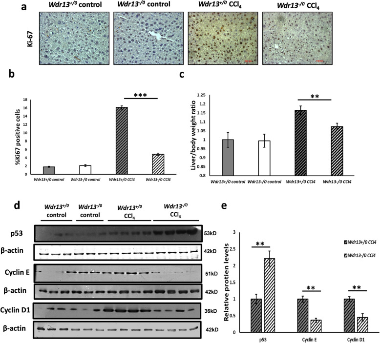
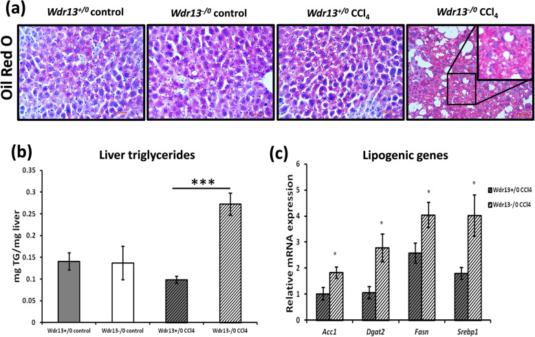
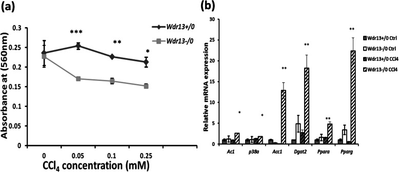
WDR13 - a WD repeat protein, is abundant in pancreas, liver, ovary and testis. Absence of this protein in mice has been seen to be associated with pancreatic β-cell proliferation, hyperinsulinemia and age dependent mild obesity. Previously, we have reported that the absence of WDR13 in diabetic Leprdb/db mice helps in amelioration of fatty liver phenotype along with diabetes and systemic inflammation. This intrigued us to study direct liver injury and hepatic regeneration in Wdr13-/0 mice using hepatotoxin CCl4. In the present study we report slower hepatic regeneration in Wdr13-/0 mice as compared to their wild type littermates after CCl4 administration. Interestingly, during the regeneration phase, hepatic hypertriglyceridemia was observed in Wdr13-/0 mice. Further analyses revealed an upregulation of PPAR pathway in the liver of CCl4- administered Wdr13-/0 mice, causing de novo lipogenesis. The slower hepatic regeneration observed in CCl4 administered Wdr13-/0 mice, may be linked to liver hypertriglyceridemia because of activation of PPAR pathway.
Keywords: Fatty liver; Hepatosteatosis; Hepatotoxin; Hypertriglyceridemia; PPARg.
Conflict of interest statement
Authors declare no conflict of interest.
Figures


Similar articles
-
Genetic deletion of Wdr13 improves the metabolic phenotype of Lepr (db/db) mice by modulating AP1 and PPARγ target genes.Diabetologia. 2015 Feb;58(2):384-92. doi: 10.1007/s00125-014-3438-y. Epub 2014 Nov 23. Diabetologia. 2015. PMID: 25417213
-
Wdr13 and streptozotocin-induced diabetes.Nutr Diabetes. 2018 Oct 29;8(1):57. doi: 10.1038/s41387-018-0065-6. Nutr Diabetes. 2018. PMID: 30369599 Free PMC article.
-
WD-repeat protein WDR13 is a novel transcriptional regulator of c-Jun and modulates intestinal homeostasis in mice.BMC Cancer. 2017 Feb 21;17(1):148. doi: 10.1186/s12885-017-3118-7. BMC Cancer. 2017. PMID: 28222755 Free PMC article.
-
Hepatotoxicity and mechanism of action of haloalkanes: carbon tetrachloride as a toxicological model.Crit Rev Toxicol. 2003;33(2):105-36. doi: 10.1080/713611034. Crit Rev Toxicol. 2003. PMID: 12708612 Review.
-
Hepatic lipid accumulation: cause and consequence of dysregulated glucoregulatory hormones.J Endocrinol. 2017 Jul;234(1):R1-R21. doi: 10.1530/JOE-16-0513. Epub 2017 Apr 20. J Endocrinol. 2017. PMID: 28428362 Review.
Cited by
-
Tcf7l2 in hepatocytes regulates de novo lipogenesis in diet-induced non-alcoholic fatty liver disease in mice.Diabetologia. 2023 May;66(5):931-954. doi: 10.1007/s00125-023-05878-8. Epub 2023 Feb 10. Diabetologia. 2023. PMID: 36759348 Free PMC article.
-
Therapeutic Effect of Polymeric Nanomicelles Formulation of LY2157299-Galunisertib on CCl4-Induced Liver Fibrosis in Rats.J Pers Med. 2022 Nov 1;12(11):1812. doi: 10.3390/jpm12111812. J Pers Med. 2022. PMID: 36579532 Free PMC article.
-
Molecular characterization of Wdr13 knockout female mice uteri: a model for human endometrial hyperplasia.Sci Rep. 2020 Sep 3;10(1):14621. doi: 10.1038/s41598-020-70773-w. Sci Rep. 2020. PMID: 32883989 Free PMC article.
References
-
- Reddy JK, Rao MS. Lipid metabolism and liver inflammation. II. Fatty liver disease and fatty acid oxidation. Am J Physiol Gastrointest Liver Physiol. 2006;290(5):G852–8. - PubMed
LinkOut - more resources
Full Text Sources
Miscellaneous
