

Cerebral sterile inflammation in neurodegenerative diseases
- PMID: 33292860
- PMCID: PMC7722432
- DOI: 10.1186/s41232-020-00137-4
Cerebral sterile inflammation in neurodegenerative diseases
Abstract
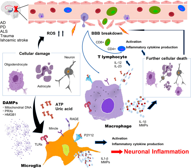
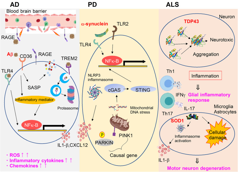
Therapeutic strategies for regulating neuroinflammation are expected in the development of novel therapeutic agents to prevent the progression of central nervous system (CNS) pathologies. An understanding of the detailed molecular and cellular mechanisms of neuroinflammation in each CNS disease is necessary for the development of therapeutics. Since the brain is a sterile organ, neuroinflammation in Alzheimer's disease (AD), Parkinson's disease (PD), and amyotrophic lateral sclerosis (ALS) is triggered by cerebral cellular damage or the abnormal accumulation of inflammatogenic molecules in CNS tissue through the activation of innate and acquired immunity. Inflammation and CNS pathologies worsen each other through various cellular and molecular mechanisms, such as oxidative stress or the accumulation of inflammatogenic molecules induced in the damaged CNS tissue. In this review, we summarize the recent evidence regarding sterile immune responses in neurodegenerative diseases.
Keywords: Alzheimer’s disease; Amyotrophic lateral sclerosis; Neuroinflammation; Parkinson’s disease.
Conflict of interest statement
The authors declare that they have no competing interests.
Figures
References
-
- Fraser PA. The role of free radical generation in increasing cerebrovascular permeability. Free Radic Biol Med. 2011;51:967–977. - PubMed
Publication types
LinkOut - more resources
Full Text Sources
Miscellaneous
