Review
. 2021 Apr:149:106212.
doi: 10.1016/j.envint.2020.106212. Epub 2020 Dec 5.

Low dose ionizing radiation effects on the immune system

Affiliations
Review

Low dose ionizing radiation effects on the immune system

Katalin Lumniczky et al. Environ Int. 2021 Apr.

Abstract

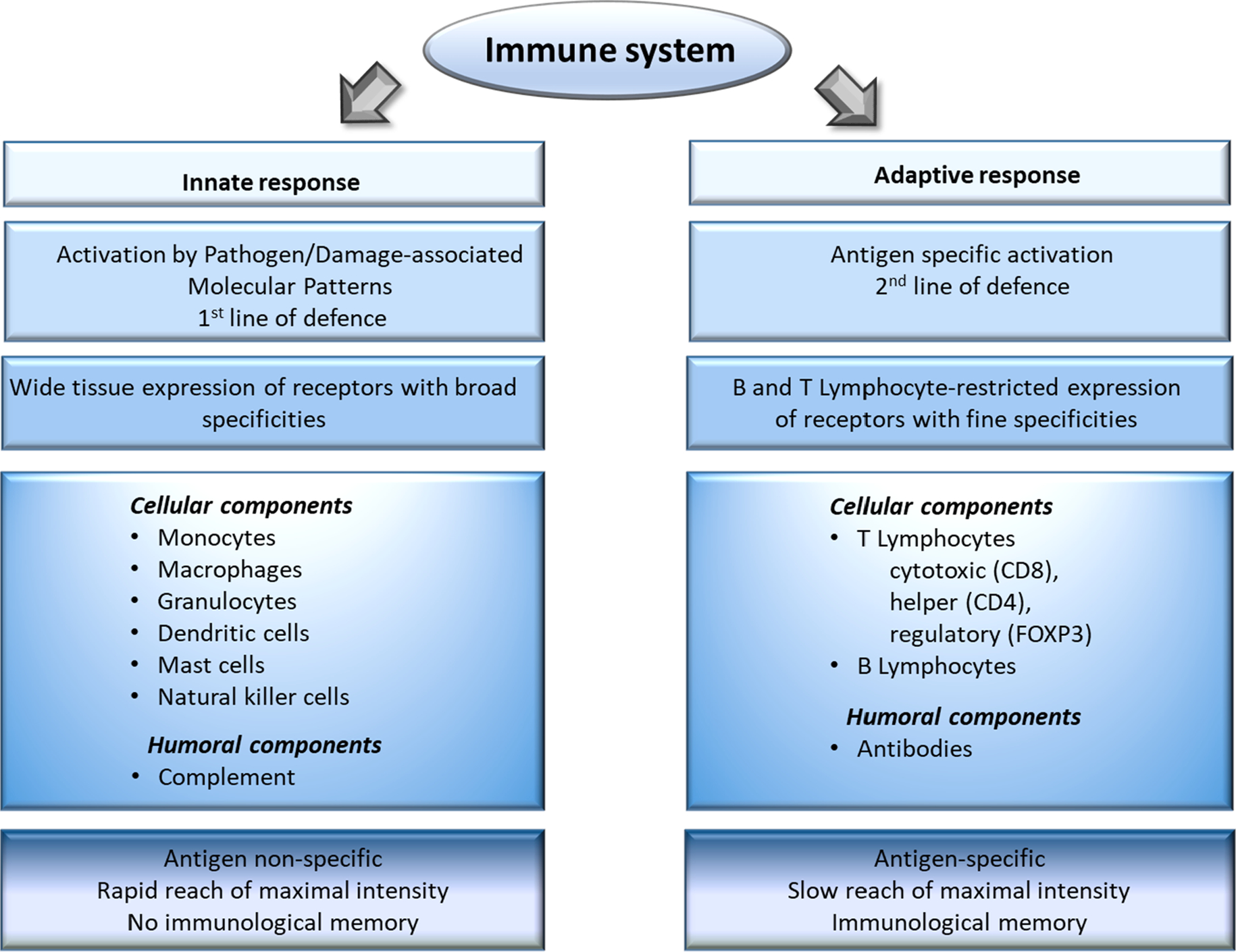
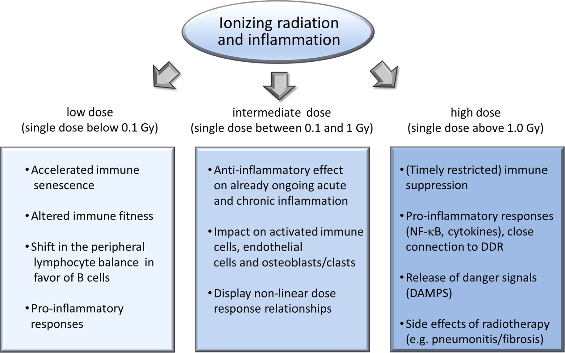
Ionizing radiation interacts with the immune system in many ways with a multiplicity that mirrors the complexity of the immune system itself: namely the need to maintain a delicate balance between different compartments, cells and soluble factors that work collectively to protect, maintain, and restore tissue function in the face of severe challenges including radiation damage. The cytotoxic effects of high dose radiation are less relevant after low dose exposure, where subtle quantitative and functional effects predominate that may go unnoticed until late after exposure or after a second challenge reveals or exacerbates the effects. For example, low doses may permanently alter immune fitness and therefore accelerate immune senescence and pave the way for a wide spectrum of possible pathophysiological events, including early-onset of age-related degenerative disorders and cancer. By contrast, the so called low dose radiation therapy displays beneficial, anti-inflammatory and pain relieving properties in chronic inflammatory and degenerative diseases. In this review, epidemiological, clinical and experimental data regarding the effects of low-dose radiation on the homeostasis and functional integrity of immune cells will be discussed, as will be the role of immune-mediated mechanisms in the systemic manifestation of localized exposures such as inflammatory reactions. The central conclusion is that ionizing radiation fundamentally and durably reshapes the immune system. Further, the importance of discovery of immunological pathways for modifying radiation resilience amongst other research directions in this field is implied.

Keywords: DNA damage response; Epidemiological data; Immune system; Inflammation; Low-dose ionizing radiation.


Conflict of interest statement

Declaration of Competing Interest

The authors declare that they have no known competing financial interests or personal relationships that could have appeared to influence the work reported in this paper.

Figures

Fig. 1.
Fig. 1.. Schematic representation of the structure of the immune system and its major functional features.
A molecule that is recognized by the immune system is called an antigen, which can be both self and non-self in origin. The immune system can be divided in two main compartments: the innate immune system and the adaptive immune system. The innate immune system is composed of a cellular compartment consisting of mononuclear cells (monocytes/macrophages, mast cells), polymorphonuclear cells (neutrophils, basophils, eosinophils), dendritic cells (DCs), innate immune cells (e.g. natural killer or NK cells) and the humoral complement system (Artis and Spits, 2015). Innate immune cells see danger through their germline-encoded pattern recognition receptors (PRRs), which recognize specific molecular structures present on pathogens (so-called pathogen-associated molecular patterns or PAMPs) or produced by damaged cells (so-called damage-associated molecular patterns or DAMPs) (Amarante-Mendes et al., 2018). Forming our first-line of defense, this recognition is relatively non-specific and quick, reaching its maximal intensity shortly after antigen encounter without yielding specific immunological memory. Phagocytosis is one of the main mechanisms for antigen elimination by innate immune cells. During danger recognition and antigen processing innate immune cells mature and release various soluble immune mediators called cytokines and chemokines, which drive inflammation and attract adaptive immune cells (Commins et al., 2010). In fact, an important role of the innate immune system is the activation of the adaptive arm. Macrophages and DCs in particular are professional antigen presenting cells with the unique ability to activate naïve cells of the adaptive immune system by displaying components of the processed antigens within the major histocompatibility complex (MHC) on their surface and present them to lymphocytes in the presence of necessary co-stimulatory signals (Wynn et al., 2013). Cells of the adaptive immune system include T lymphocytes (such as CD4 + helper, CD8 + cytotoxic and Foxp3 + regulatory) and B lymphocytes. T cells are responsible for cell-mediated immune response while B cells play role in humoral immune response (mediated by antibodies). In contrast to the innate immune system, the major features of the adaptive immune response are: high antigen specificity, latency of maximal response and development of immunological memory exemplified by faster and qualitatively different recall responses (Santana and Esquivel-Guadarrama, 2006). The first step in the activation of the adaptive immune system is antigen recognition by CD4 + or CD8 + cells through their highly antigen-specific T-cell receptors (TCRs). Professional antigen presenting cells present antigenic peptides conjugated either to MHCII, inducing CD4 + activation or to MHCI, contributing to CD8 + activation. Activated CD8 + T cells kill the antigen-presenting cells through the release of cytotoxic agents stored in intracellular granules, or directly by cell-to-cell contact engaging death receptors, or through the production of cytokines that trigger apoptosis. B cells, on the other hand, recognize extracellular antigens via their antigen-specific B cell receptor, which are essentially antibodies bound on the cell membrane forming a transmembrane receptor. Once activated with help from CD4 + T cells, B cells start to divide and differentiate into plasma cells which secrete huge numbers of soluble antibodies similar to the one that recognized the antigen in the first place (Hardy and Hayakawa, 2001). Circulating antibodies bind to their specific antigens and these antigen–antibody complexes induce activation of the complement system, which in turn leads to a rapid neutralisation by the proteolytic activity of the complement system and further phagocytosis by innate cells, i.e. antibody-dependent cellular cytotoxicity. Most of the intercellular communication in the immune system is guided through a complex system of chemokines, cytokines and interferons that affect trafficking, activation, differentiation and functional maturation (Turner et al., 2014). To prevent tissue damage from excessive immune activation multiple control mechanisms are in place that act through cell-to-cell contact or cytokines, involving among others regulatory T cells (Tregs) (Persa et al., 2015). Finally, to mount an effective response, immune components must circulate between the blood and lymph nodes, recognize antigens upon contact with presenting cells, and differentiate to effector T cells and plasma cells. Moreover, these cells must extravasate the lymph nodes, migrate to affected tissue to secure host-protective activities and to recircle to blood to counteract chronic activation (Germain et al., 2012). Accordingly, one has to consider a high degree of cellular motility and interaction dynamics of the immune system.
Fig. 2.
Fig. 2.
Schematic representation of the most important immune- and inflammation-related processes developing after low, intermediate and high dose irradiation based on available epidemiological, clinical and experimental data.

References

    1. Abuodeh Y, Venkat P, Kim S, 2016. Systematic review of case reports on the abscopal effect. Curr. Probl. Cancer 40, 25–37. 10.1016/j.currproblcancer.2015.10.001. - DOI - PubMed
    1. Agate L, Mariotti S, Elisei R, Mossa P, Pacini F, Molinaro E, Grasso L, Masserini L, Mokhort T, Vorontsova T, Arynchyn A, Tronko MD, Tsyb A, Feldt-Rasmussen U, Juul A, Pinchera A, 2008. Thyroid autoantibodies and thyroid function in subjects exposed to Chernobyl fallout during childhood: evidence for a transient radiation-induced elevation of serum thyroid antibodies without an increase in thyroid autoimmune disease. J. Clin. Endocrinol. Metab. 93, 2729–2736. 10.1210/jc.2008-0060. - DOI - PubMed
    1. Aggarwal BB, Vijayalekshmi RV, Sung B, 2009. Targeting inflammatory pathways for prevention and therapy of cancer: short-term friend, long-term foe. Clin. Cancer Res. 15, 425–430. 10.1158/1078-0432.CCR-08-0149. - DOI - PubMed
    1. Ahmad IM, Temme JB, Abdalla MY, Zimmerman MC, 2016. Redox status in workers occupationally exposed to long-term low levels of ionizing radiation: A pilot study. Redox Rep. 21, 139–145. 10.1080/13510002.2015.1101891. - DOI - PMC - PubMed
    1. Akiyama M, 1995. Late effects of radiation on the human immune system: an overview of immune response among the atomic-bomb survivors. Int. J. Radiat Biol. 68, 497–508. 10.1080/09553009514551491. - DOI - PubMed

Publication types