Case Reports
. 2020 Dec 31;36(4):258-262.
doi: 10.5758/vsi.200042.

Hybrid Treatment of Aberrant Right Subclavian Artery Causing Dysphagia Lusoria by Subclavian to Carotid Transposition and Endovascular Plug

Affiliations
Case Reports

Hybrid Treatment of Aberrant Right Subclavian Artery Causing Dysphagia Lusoria by Subclavian to Carotid Transposition and Endovascular Plug

Monica Leon et al. Vasc Specialist Int. .

Abstract

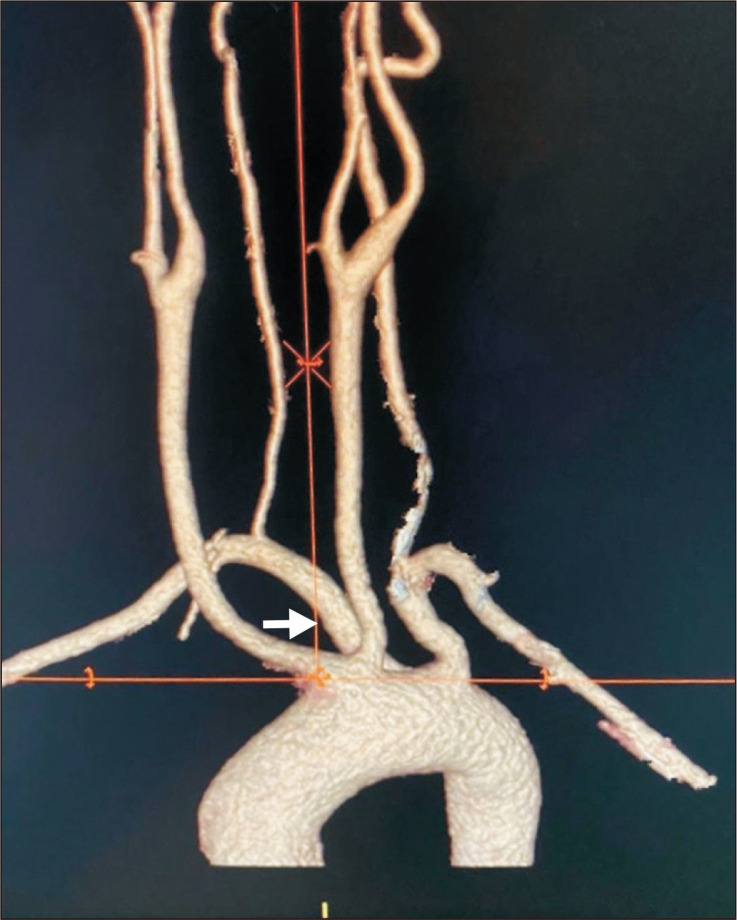
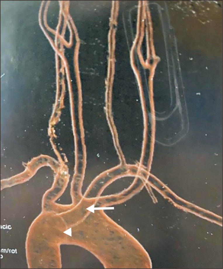
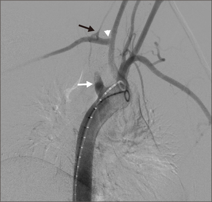
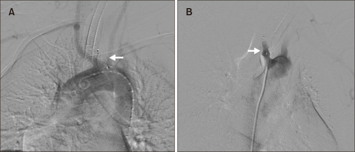
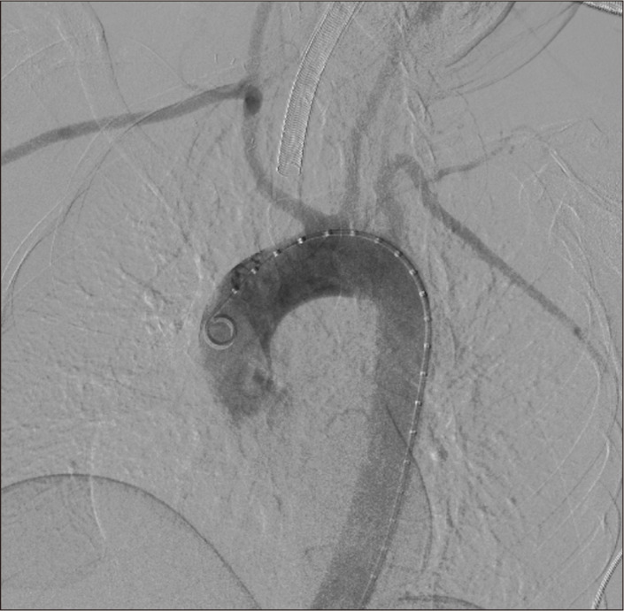
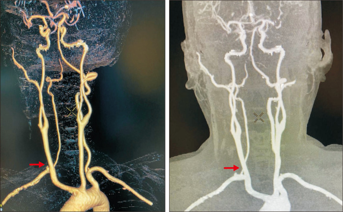
Differences in the common aortic arch branching pattern arise from abnormal embryological development. Aberrant origin of the right subclavian artery is the most common of these anomalies. We report the case of a 47-year-old female with progressive dysphagia, found to have an aberrant right subclavian artery (ARSA) running posterior to the esophagus on computed tomography angiography. She was managed successfully with a hybrid procedure involving a right supraclavicular incision for ARSA ligation and subclavian to carotid transposition followed by endovascular closure of the ARSA origin.

Keywords: Cardiovascular abnormalities; Endovascular procedures; Subclavian artery.


Conflict of interest statement

CONFLICTS OF INTEREST

The authors have nothing to disclose.

Figures

Fig. 1
Fig. 1
Preoperative three-dimensional reconstruction of computed tomography scan showed an aberrant right subclavian artery (arrow).
Fig. 2
Fig. 2
Posterior view of preoperative computed tomography scan showed aneurysmal degeneration at the origin (arrow head) of the aberrant right subclavian artery (arrow).
Fig. 3
Fig. 3
Axial view of computed tomography scan showed: (A) The origin of the aberrant right subclavian artery (arrow). (B) Posterior to the esophagus with esophageal compression (arrow).
Fig. 4
Fig. 4
Angiogram (right anterior oblique) showed the ab- errant right subclavian artery origin to have a dilatation (white arrow), subclavian to carotid transposition (arrow head), and vertebral artery preservation (black arrow).
Fig. 5
Fig. 5
Left anterior oblique view (A) and right anterior oblique view (B) of angiogram showed endovascular closure of the aberrant right subclavian artery stump with Amplatzer vascular plug (arrows).
Fig. 6
Fig. 6
Completion angiogram showed patent right subclavian to carotid transposition with absence of contrast material in the aberrant right subclavian artery origin.
Fig. 7
Fig. 7
Six-month follow-up computed tomography angiography of the neck and chest showed a patent right subclavian to carotid transposition and vertebral artery preservation (arrows).

References

    1. Jakanani GC, Adair W. Frequency of variations in aortic arch anatomy depicted on multidetector CT. Clin Radiol. 2010;65:481–487. doi: 10.1016/j.crad.2010.02.003. - DOI - PubMed
    1. Popieluszko P, Henry BM, Sanna B, Hsieh WC, Saganiak K, Pękala PA, et al. A systematic review and meta-analysis of variations in branching patterns of the adult aortic arch. J Vasc Surg. 2018;68:298–306.e10. doi: 10.1016/j.jvs.2017.06.097. - DOI - PubMed
    1. Bennett AL, Cock C, Heddle R, Morcom RK. Dysphagia lusoria: a late onset presentation. World J Gastroenterol. 2013;19:2433–2436. doi: 10.3748/wjg.v19.i15.2433. - DOI - PMC - PubMed
    1. Baker AC, Atkins BZ, Clouse WD, Noll R, Sampson J, Williams T. Repair of aberrant right subclavian artery entirely via a supraclavicular approach. Ann Vasc Surg. 2014;28:489.e1–489.e4. doi: 10.1016/j.avsg.2013.11.001. - DOI - PubMed
    1. Felson B, Cohen S, Courter SR, McGuire J. Anomalous right subclavian artery. Radiology. 1950;54:340–349. doi: 10.1148/54.3.340. - DOI - PubMed

Publication types

LinkOut - more resources