. 2019 Dec 10:7:36.
doi: 10.1038/s41413-019-0075-6. eCollection 2019.

Mesenchymal VEGFA induces aberrant differentiation in heterotopic ossification

Affiliations

Mesenchymal VEGFA induces aberrant differentiation in heterotopic ossification

Charles Hwang et al. Bone Res. .

Abstract

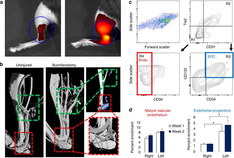
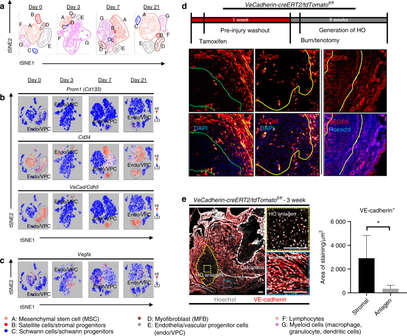
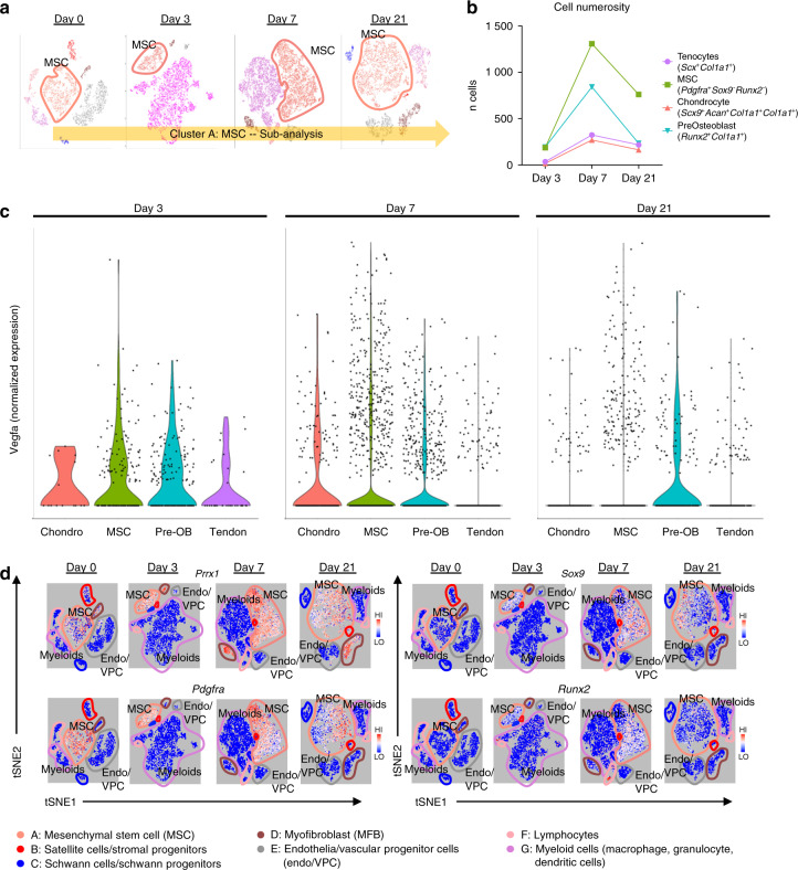
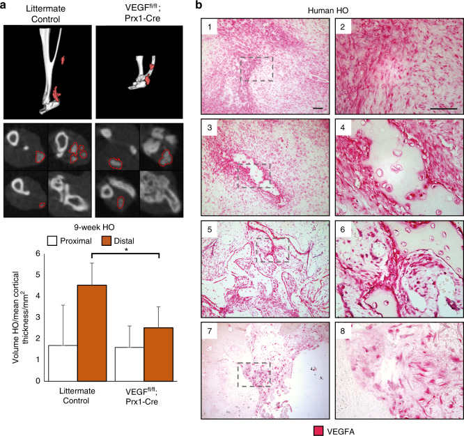
Heterotopic ossification (HO) is a debilitating condition characterized by the pathologic formation of ectopic bone. HO occurs commonly following orthopedic surgeries, burns, and neurologic injuries. While surgical excision may provide palliation, the procedure is often burdened with significant intra-operative blood loss due to a more robust contribution of blood supply to the pathologic bone than to native bone. Based on these clinical observations, we set out to examine the role of vascular signaling in HO. Vascular endothelial growth factor A (VEGFA) has previously been shown to be a crucial pro-angiogenic and pro-osteogenic cue during normal bone development and homeostasis. Our findings, using a validated mouse model of HO, demonstrate that HO lesions are highly vascular, and that VEGFA is critical to ectopic bone formation, despite lacking a contribution of endothelial cells within the developing anlagen.

Keywords: Bone; Pathogenesis.


Conflict of interest statement

Competing interestsA.W.J. serves on the scientific advisory board for Novadip LLC and also receives laboratory financial support from MTF Biologics for research unrelated to the current project. All other authors declare no competing interests..

Figures

Fig. 1
Fig. 1
Focal injury superimposed upon systemic inflammation induces robust local angiogenesis and produces enrichment of endothelial progenitors at the injury site. a Angiosense IVIS demonstrates increased perfusion to injured hindlimb at 20 h post surgery (representative images from n = 3 per group). b MICROFIL perfusion nano-CT images and micro-CT of mature mice show networks of nascent vessel outgrowth in regions of subsequent HO formation following burn tenotomy (green inset) at 9 weeks. Vessels in the distal hindlimb exhibit dense vascular infiltration (red inset). c FACS gating schema of injury site 2 weeks after burn tenotomy. CD31 Tie2 CD34+ CD133+ endothelial progenitor cells (EPC, blue) identified via R1 + R3. CD31+ Tie2+ CD34 mature endothelial cells (red) identified via R2 + R4. Remaining live events were collected by surface marker depletion. d Proportion of gated EPC and mature endothelium from all live cells as assessed by flow cytometry was quantified at 1 week and 2 weeks post burn tenotomy
Fig. 2
Fig. 2
Vascular progenitors are not the source for VEGFA in HO formation. a Resultant seven clusters identifying cellular subpopulations within HO anlagen at respective timepoints. b Expression profiles for vascular progenitor genes Prom1 (Cd133), CD34, and Vecad/Cdh5. c Expression profile for Vegfa d Schematic for induction protocol of endogenous reporter mice. VeCadherin-creERT2/tdTomatofl/fl identify cells with endothelial programming within outlined regions. Sections taken from distal hindlimb of injury. Right: Sections of injury site from wild-type 3 week burn/tenotomy mice labeled for VEGFA. e Tiled immunofluorescent micrograph of 3 week burn/tenotomy injury site in endogenous VeCadherin reporters. Quantifications for thresholded total positive area (FIJI/ImageJ) were performed across biological triplicate, with 2 hpf per specimen for both HO anlagen site vs. surrounding stroma (n = 6/group)
Fig. 3
Fig. 3
Mesenchymal progenitor cells comprise the majority of VEGFA expressing cells within HO lesions. Clusters from 10× RNA sequencing across days 0, 3, 7, and 21 timepoints. Expression profiles corresponding to a mesenchymal progenitor cells: Pdgfra, Osr2, and En1; b Vegfa; c and macrophages: Mrc1 and Adgre1. d Expression heatmap delineating timecourse for log-fold changes in expression for genes of interest within (Top) mesenchymal stem cell cluster A and (Bottom) endothelial cells/vascular progenitor cells cluster E. Red and blue signify upregulation and downregulation respectively. e Representative immunolabeling from burn/tenotomy on wild-type mice 1 week after injury. n = 3/group. Color dotted squares designate magnified regions. White dotted lines circumscribe residual bellies of Achilles tendon. Dash dot lines outline reference native bone (calcaneus). Scalebars represent 100 μm. f Quantifications for relative signal in 1-week injury site (left leg) vs. uninjured contralateral limb (right leg) following automated thresholding (Yen). Linear regression of PDGFRα vs. VEGFA demonstrates significant correlation
Fig. 4
Fig. 4
Vegfa is variably expressed amongst MSC progenitors and their differentiated progeny. a Schematic of cluster subanalysis of MSC cells from single-cell RNA sequencing. b Discrete counts of cell types and classification criteria. c Expression levels of Vegfa across all indicated cell types. d Expression profiles of characteristic markers Prrx1; mesenchymal progenitors: Pdgfra; chondrocytes: Sox9; and preosteoblasts: Runx2
Fig. 5
Fig. 5
VEGFA knockout and molecular inhibition attenuates formation of HO in the distal hindlimb. Following 9 weeks burn tenotomy, a Prx-Cre;Vegffl/fl; treated WT mice exhibit lower volumes of heterotopic ossification than their corresponding controls at 800 HU. Prx-Cre HO volumes normalized by mean cortical thickness (mm) due to congenital phenotype. n = 4–9/group. Bars represent means and SD. *P < 0.05 vs. Control by Student’s t test. b Magnified images of human HO samples immunolabeled for VEGFA: (1,2) pre-HO stroma, (3,4) early HO formation with surrounding stromal fibroblasts, (5,6) intermediate HO, and (7,8) mature HO with terminal ossification. VEGFA indicated by red, scalebar indicates 100 μm

References

    1. Ranganathan K, et al. Heterotopic ossification: basic-science principles and clinical correlates. J. Bone Jt. Surg. Am. 2015;97:1101–1111. doi: 10.2106/JBJS.N.01056. - DOI - PMC - PubMed
    1. Potter BK, Burns TC, Lacap AP, Granville RR, Gajewski DA. Heterotopic ossification following traumatic and combat-related amputations. Prevalence, risk factors, and preliminary results of excision. J. Bone Jt. Surg. Am. 2007;89:476–486. doi: 10.2106/00004623-200703000-00003. - DOI - PubMed
    1. Ring D, Jupiter JB. Excision of heterotopic bone around the elbow. Tech. Hand Up. Extrem Surg. 2004;8:25–33. doi: 10.1097/00130911-200403000-00007. - DOI - PubMed
    1. Pavey GJ, et al. What risk factors predict recurrence of heterotopic ossification after excision in combat-related amputations? Clin. Orthop. Relat. Res. 2015;473:2814–2824. doi: 10.1007/s11999-015-4266-1. - DOI - PMC - PubMed
    1. Genet F, et al. The impact of preoperative hip heterotopic ossification extent on recurrence in patients with head and spinal cord injury: a case control study. PloS ONE. 2011;6:e23129. doi: 10.1371/journal.pone.0023129. - DOI - PMC - PubMed