Meta-Analysis
. 2020 Dec 8;11(1):6285.
doi: 10.1038/s41467-020-19111-2.

Cerebral small vessel disease genomics and its implications across the lifespan

Muralidharan Sargurupremraj  1 Hideaki Suzuki  2   3   4 Xueqiu Jian  5   6 Chloé Sarnowski  7 Tavia E Evans  8   9 Joshua C Bis  10 Gudny Eiriksdottir  11 Saori Sakaue  12   13   14 Natalie Terzikhan  15 Mohamad Habes  6   16   17 Wei Zhao  18 Nicola J Armstrong  19 Edith Hofer  20   21 Lisa R Yanek  22 Saskia P Hagenaars  23   24 Rajan B Kumar  25 Erik B van den Akker  26   27   28 Rebekah E McWhirter  29   30 Stella Trompet  31   32 Aniket Mishra  1 Yasaman Saba  1   33 Claudia L Satizabal  6   34   35 Gregory Beaudet  36 Laurent Petit  36 Ami Tsuchida  36 Laure Zago  36 Sabrina Schilling  1 Sigurdur Sigurdsson  11 Rebecca F Gottesman  37 Cora E Lewis  38 Neelum T Aggarwal  39 Oscar L Lopez  40 Jennifer A Smith  18   41 Maria C Valdés Hernández  23   42   43 Jeroen van der Grond  44 Margaret J Wright  45   46 Maria J Knol  15 Marcus Dörr  47   48 Russell J Thomson  30   49 Constance Bordes  1 Quentin Le Grand  1 Marie-Gabrielle Duperron  1 Albert V Smith  11 David S Knopman  50 Pamela J Schreiner  51 Denis A Evans  52 Jerome I Rotter  53 Alexa S Beiser  7   34   35 Susana Muñoz Maniega  23   42 Marian Beekman  26 Julian Trollor  54   55 David J Stott  56 Meike W Vernooij  9   15 Katharina Wittfeld  57 Wiro J Niessen  9   58 Aicha Soumaré  1 Eric Boerwinkle  59 Stephen Sidney  60 Stephen T Turner  61 Gail Davies  21   62 Anbupalam Thalamuthu  53 Uwe Völker  63 Mark A van Buchem  43 R Nick Bryan  64 Josée Dupuis  6   32 Mark E Bastin  21   41 David Ames  65   66 Alexander Teumer  15   47 Philippe Amouyel  67   68 John B Kwok  69   70 Robin Bülow  71 Ian J Deary  21   62 Peter R Schofield  70   72 Henry Brodaty  53   73 Jiyang Jiang  53 Yasuharu Tabara  74 Kazuya Setoh  75 Susumu Miyamoto  75 Kazumichi Yoshida  75 Manabu Nagata  75 Yoichiro Kamatani  76 Fumihiko Matsuda  74 Bruce M Psaty  77   78 David A Bennett  79 Philip L De Jager  80   81 Thomas H Mosley  82 Perminder S Sachdev  53   83 Reinhold Schmidt  18 Helen R Warren  84   85 Evangelos Evangelou  86   87 David-Alexandre Trégouët  1 International Network against Thrombosis (INVENT) ConsortiumInternational Headache Genomics Consortium (IHGC)Mohammad A Ikram  15 Wei Wen  54 Charles DeCarli  88 Velandai K Srikanth  30   89 J Wouter Jukema  32 Eline P Slagboom  26 Sharon L R Kardia  18 Yukinori Okada  12   13   90 Bernard Mazoyer  36 Joanna M Wardlaw  23   42   43   91 Paul A Nyquist  92   93 Karen A Mather  54   72 Hans J Grabe  94   95 Helena Schmidt  33 Cornelia M Van Duijn  96 Vilmundur Gudnason  11   97 William T Longstreth Jr  98 Lenore J Launer  99   100 Mark Lathrop  101 Sudha Seshadri  6   34   35 Christophe Tzourio  1   102 Hieab H Adams  8   9 Paul M Matthews  4   103   104 Myriam Fornage  105 Stéphanie Debette  106   107   108
Collaborators, Affiliations
Meta-Analysis

Cerebral small vessel disease genomics and its implications across the lifespan

Muralidharan Sargurupremraj et al. Nat Commun. .

Abstract

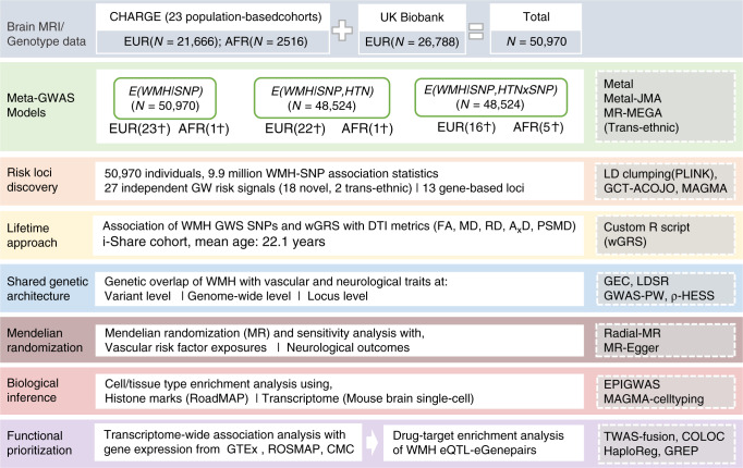
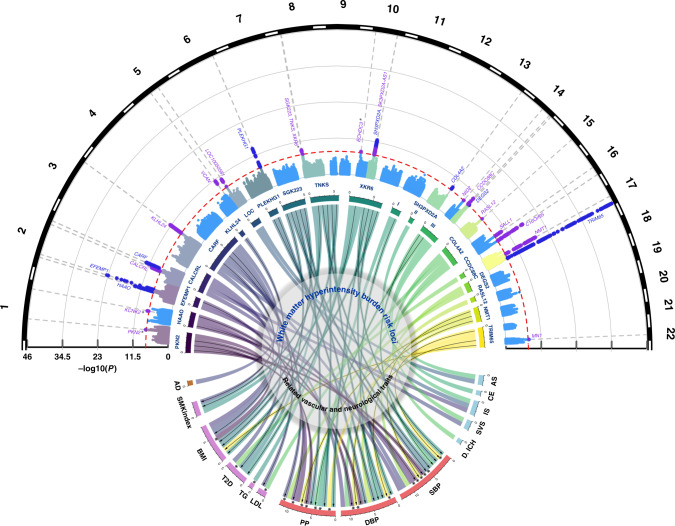
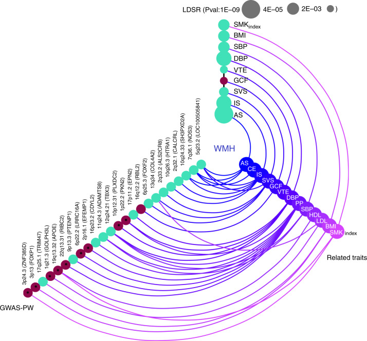
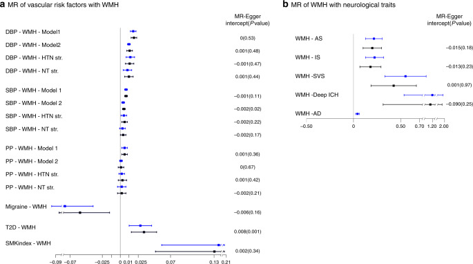
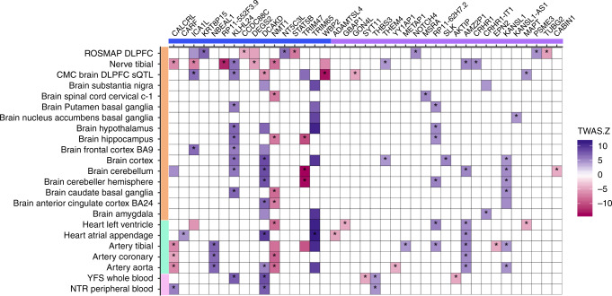
White matter hyperintensities (WMH) are the most common brain-imaging feature of cerebral small vessel disease (SVD), hypertension being the main known risk factor. Here, we identify 27 genome-wide loci for WMH-volume in a cohort of 50,970 older individuals, accounting for modification/confounding by hypertension. Aggregated WMH risk variants were associated with altered white matter integrity (p = 2.5×10⁻⁷) in brain images from 1,738 young healthy adults, providing insight into the lifetime impact of SVD genetic risk. Mendelian randomization suggested causal association of increasing WMH-volume with stroke, Alzheimer-type dementia, and of increasing blood pressure (BP) with larger WMH-volume, notably also in persons without clinical hypertension. Transcriptome-wide colocalization analyses showed association of WMH-volume with expression of 39 genes, of which four encode known drug targets. Finally, we provide insight into BP-independent biological pathways underlying SVD and suggest potential for genetic stratification of high-risk individuals and for genetically-informed prioritization of drug targets for prevention trials.


Conflict of interest statement

The authors declare no competing interests.

Figures

Fig. 1
Fig. 1. Study workflow and rationale.
Ϯ number of GW hits. MRI magnetic resonance imaging, CHARGE cohorts for heart and aging research in genomic epidemiology, EUR European, AFR African–american, GWAS genome-wide association study, WMH White matter hyperintensities, SNP single nucleotide polymorphism, HTN hypertension, JMA joint meta-analysis, MR-MEGA meta-regression of multi-ethnic genetic association, GW genome-wide, LD linkage disequilibrium, GCTA-COJO genome-wide complex trait analysis- conditional and joint analysis, MAGMA multi-marker analysis of genomic annotation, DTI diffusion tensor imaging, iSHARE internet based student health research enterprise, FA fractional anisotropy, MD mean diffusivity, RD radial diffusivity, AxD axial diffusivity, PSMD peak width of skeletonised mean diffusivity, wGRS weighted genetic risk score, GEC genetic type I error calculator, LDSR LD-score regression, GWAS-PW GWAS-pairwise analysis, HESS heritability estimator from summary statistics, EPIGWAS epigenome wide association study, TWAS transcriptome-wide association study, GTEx genotype-tissue expression, ROSMAP religious orders study and the RUSH memory and aging project, CMC common mind consortium, eQTL expression quantitative trait loci, eGene expression-associated genes, COLOC colocalisation, GREP genome for repositioning drugs.
Fig. 2
Fig. 2. Genome-wide association results with WMH burden and genetic overlap of WMH risk loci.
Circular Manhattan plot (top) displaying novel (violet) and known (dark blue) genome-wide significant WMH risk loci (dotted line: P < 5 × 10−8). Asterisks denote association signals that reach genome-wide significance only in the HTN-adjusted model (PKN2, XKR6) or in the MR-MEGA transethnic meta-analysis (KCNK2, ECHDC3). Chord diagram (center) summarizing the association of genome-wide significant risk variants for WMH burden (upper section) with vascular and neurological traits (bottom section) (P < 1.3 × 10−4, see Methods). The width of each of the stems corresponds to the number of traits associated with a given locus (upper section) or the number of loci associated with a given trait. Black arrows indicate genome-wide significant associations, and asterisks denote SNPs exhibiting unexpected directionality of associations (WMH risk allele displaying protective association with vascular or neurological traits). LOC* LOC100505841, SBP systolic blood pressure, DBP diastolic blood pressure, PP pulse pressure, AS all stroke, IS ischemic stroke, SVS small vessel stroke, CE cardioembolic stroke, ICH intracerebral hemorrhage, AD Alzheimer’s disease, BMI body mass index, LDL low-density lipoprotein, VTE venous thromboembolism, T2D type II diabetes, MIG migraine, SMKindex lifetime smoking index, WMH white matter hyperintensity.
Fig. 3
Fig. 3. Shared genetic architecture of WMH at genome-wide and regional level Color coded for the direction of effect (Green: Positive genetic correlation; Red: Negative genetic correlation).
The LD-score regression (LDSR) axis shows evidence for genome-wide correlations (after Bonferroni correction for multiple testing P < 3.6 × 10−3, Methods), with the size of the nodes corresponding to the level of significance of the association. The GWAS-pairwise (PW) axis shows evidence for regional level overlap of association signals between WMH burden and related vascular and neurological traits (PPA3 ≥ 0.90, Methods). For any given region, the nearest gene (in brackets) to the top SNP associated with WMH is shown. Bivariate heritability estimator from summary statistics (ρ-HESS) was used to infer directionality of shared association signals (Methods) and asterisks denote an unexpected directionality of association. SBP systolic blood pressure, DBP diastolic blood pressure, PP pulse pressure, AS all stroke, IS ischemic stroke, SVS small vessel stroke, CE cardioembolic stroke, BMI body mass index, HDL high-density lipoprotein, LDL low-density lipoprotein, VTE venous thromboembolism, GCF general cognitive function, SMKindex lifetime smoking index, WMH white matter hyperintensity.
Fig. 4
Fig. 4. Mendelian randomization results of vascular risk factors with WMH burden (box A) and WMH burden with neurological traits (box B).
Point estimates and confidence intervals (blue) from the inverse-variance weighted (IVW) method are shown along with the point estimates and 95% confidence interval (black) from sensitivity analyses after filtering out potentially pleiotropic outlier variants. The intercept and p-value from the MR-Egger method is displayed on the far right (an intercept term significantly differing from zero at the conservative threshold of P < 0.05 suggests the presence of directional pleiotropy). SBP systolic blood pressure, DBP diastolic blood pressure, PP pulse pressure, HTN hypertensive, NT normotensive, Str. stratum, AS all stroke, IS ischemic stroke, SVS small vessel stroke, ICH intracerebral hemorrhage, AD Alzheimer’s disease, T2D type II diabetes, SMKindex lifetime smoking index, WMH white matter hyperintensity, Model 1 Main effects adjusted for age, sex, principal components for population stratification, intracranial volume; Model 2 Model 1 + hypertension status.
Fig. 5
Fig. 5. Transcriptome-wide association study of WMH and gene-expression datasets.
Only genes showing significant colocalization between the eQTL and the WMH risk variant in at least one tissue are shown. Susceptibility genes are depicted on the x-axis (blue: known; violet: novel), with tissue types of gene-expression datasets on the y-axis (orange: brain or peripheral nerve tissue; green: arterial/heart; pink: blood). Blue boxes correspond to WMH risk alleles being associated with upregulation (+) of gene expression in the corresponding tissues, while red boxes correspond to WMH risk alleles being associated with downregulation (−) of gene expression (color intensity corresponds to the magnitude of gene-expression effect size). Only significant TWAS associations at P < 1.1 × 10−5 are shown. Asterisks denote loci harboring a common causal variant associated with WMH and gene expression with high posterior probability using colocalization analyses (Methods; PP4 ≥ 0.75). ROSMAP religious order study and rush memory and aging project, DLPFC dorsolateral prefrontal cortex, CMC common mind consortium, BA Brodmann area, YFS young Finns study, NTR Netherlands twins register.

References

    1. Debette S, Markus HS. The clinical importance of white matter hyperintensities on brain magnetic resonance imaging: systematic review and meta-analysis. BMJ. 2010;341:c3666. doi: 10.1136/bmj.c3666. - DOI - PMC - PubMed
    1. Shi Y, et al. Cerebral blood flow in small vessel disease: a systematic review and meta-analysis. J. Cereb. Blood Flow. Metab. 2016;36:1653–1667. doi: 10.1177/0271678X16662891. - DOI - PMC - PubMed
    1. Debette S, Schilling S, Duperron MG, Larsson SC, Markus HS. Clinical significance of magnetic resonance imaging markers of vascular brain injury: a systematic review and meta-analysis. JAMA Neurol. 2019;76:81–94. doi: 10.1001/jamaneurol.2018.3122. - DOI - PMC - PubMed
    1. Corriveau RA, et al. Alzheimer’s disease-related Dementias Summit 2016: National research priorities. Neurology. 2017;89:2381–2391. doi: 10.1212/WNL.0000000000004717. - DOI - PMC - PubMed
    1. The Lancet, N. Vascular disease and neurodegeneration: advancing together. Lancet Neurol. 2017;16:333. doi: 10.1016/S1474-4422(17)30086-8. - DOI - PubMed

Publication types

MeSH terms

Grants and funding