

Combinatorial Atoh1 and Gfi1 induction enhances hair cell regeneration in the adult cochlea
- PMID: 33293609
- PMCID: PMC7722738
- DOI: 10.1038/s41598-020-78167-8
Combinatorial Atoh1 and Gfi1 induction enhances hair cell regeneration in the adult cochlea
Abstract
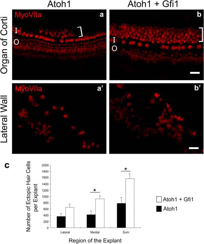
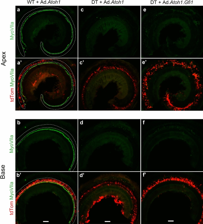
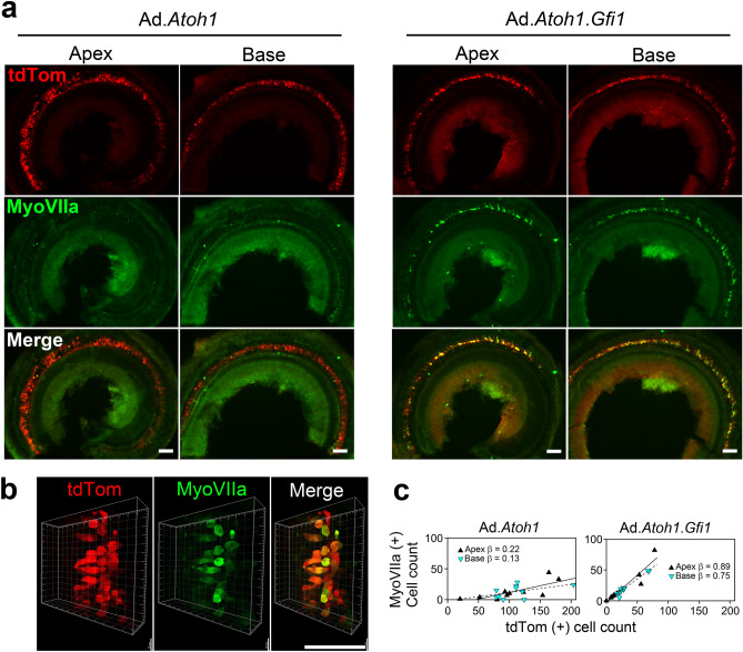
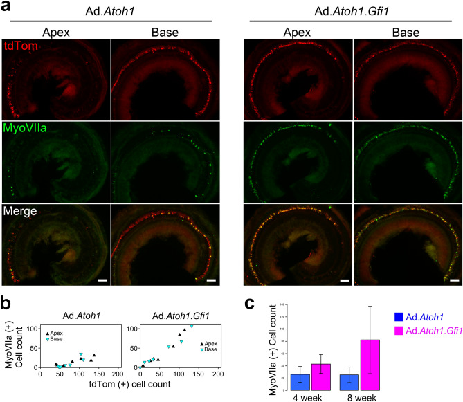
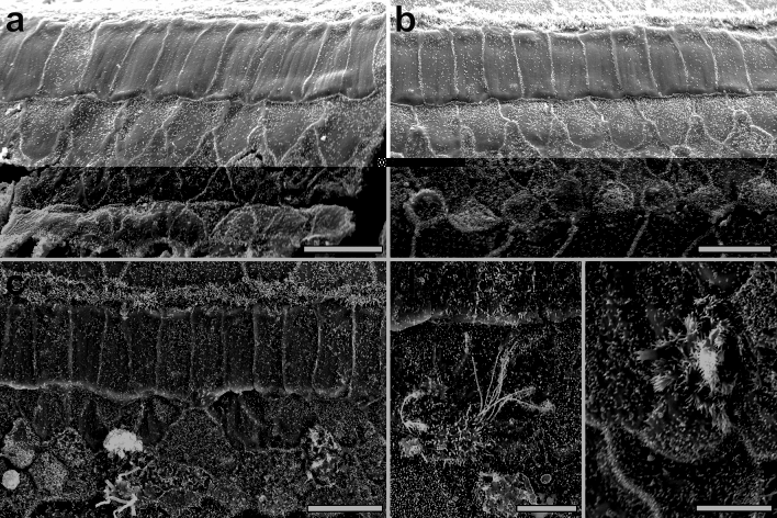
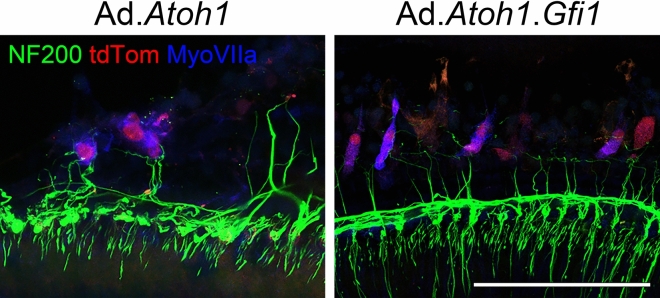
Mature mammalian cochlear hair cells (HCs) do not spontaneously regenerate once lost, leading to life-long hearing deficits. Attempts to induce HC regeneration in adult mammals have used over-expression of the HC-specific transcription factor Atoh1, but to date this approach has yielded low and variable efficiency of HC production. Gfi1 is a transcription factor important for HC development and survival. We evaluated the combinatorial effects of Atoh1 and Gfi1 over-expression on HC regeneration using gene transfer methods in neonatal cochlear explants, and in vivo in adult mice. Adenoviral over-expression of Atoh1 and Gfi1 in cultured neonatal cochlear explants resulted in numerous ectopic HC-like cells (HCLCs), with significantly more cells in Atoh1 + Gfi1 cultures than Atoh1 alone. In vitro, ectopic HCLCs emerged in regions medial to inner HCs as well as in the stria vascularis. In vivo experiments were performed in mature Pou4f3DTR mice in which HCs were completely and specifically ablated by administration of diphtheria toxin. Adenoviral expression of Atoh1 or Atoh1 + Gfi1 in cochlear supporting cells induced appearance of HCLCs, with Atoh1 + Gfi1 expression leading to 6.2-fold increase of new HCLCs after 4 weeks compared to Atoh1 alone. New HCLCs were detected throughout the cochlea, exhibited immature stereocilia and survived for at least 8 weeks. Combinatorial Atoh1 and Gfi1 induction is thus a promising strategy to promote HC regeneration in the mature mammalian cochlea.
Conflict of interest statement
The authors declare no competing interests.
Figures
Similar articles
-
Generation of mature and functional hair cells by co-expression of Gfi1, Pou4f3, and Atoh1 in the postnatal mouse cochlea.Cell Rep. 2021 Apr 20;35(3):109016. doi: 10.1016/j.celrep.2021.109016. Cell Rep. 2021. PMID: 33882317
-
Co-overexpression of Atoh1, Pou4f3, and Gfi1 enhances the transdifferentiation of supporting cells into hair cells in the neonatal mouse utricle.Neurosci Lett. 2025 Feb 16;849:138136. doi: 10.1016/j.neulet.2025.138136. Epub 2025 Jan 28. Neurosci Lett. 2025. PMID: 39884380
-
AAV-mediated Gene Cocktails Enhance Supporting Cell Reprogramming and Hair Cell Regeneration.Adv Sci (Weinh). 2024 Aug;11(29):e2304551. doi: 10.1002/advs.202304551. Epub 2024 May 29. Adv Sci (Weinh). 2024. PMID: 38810137 Free PMC article.
-
Atoh1 and other related key regulators in the development of auditory sensory epithelium in the mammalian inner ear: function and interplay.Dev Biol. 2019 Feb 15;446(2):133-141. doi: 10.1016/j.ydbio.2018.12.025. Epub 2018 Dec 31. Dev Biol. 2019. PMID: 30605626 Review.
-
Atoh1 in sensory hair cell development: constraints and cofactors.Semin Cell Dev Biol. 2017 May;65:60-68. doi: 10.1016/j.semcdb.2016.10.003. Epub 2016 Oct 14. Semin Cell Dev Biol. 2017. PMID: 27751776 Review.
Cited by
-
Ontogeny of cellular organization and LGR5 expression in porcine cochlea revealed using tissue clearing and 3D imaging.iScience. 2022 Jun 30;25(8):104695. doi: 10.1016/j.isci.2022.104695. eCollection 2022 Aug 19. iScience. 2022. PMID: 35865132 Free PMC article.
-
Expression of Atoh1, Gfi1, and Pou4f3 in the mature cochlea reprograms nonsensory cells into hair cells.Proc Natl Acad Sci U S A. 2024 Jan 30;121(5):e2304680121. doi: 10.1073/pnas.2304680121. Epub 2024 Jan 24. Proc Natl Acad Sci U S A. 2024. PMID: 38266052 Free PMC article.
-
The regenerative capacity of neonatal tissues.Development. 2022 Jun 15;149(12):dev199819. doi: 10.1242/dev.199819. Epub 2022 Jun 16. Development. 2022. PMID: 35708609 Free PMC article. Review.
-
Chromodomain helicase DNA binding protein 4 in cell fate decisions.Hear Res. 2023 Sep 1;436:108813. doi: 10.1016/j.heares.2023.108813. Epub 2023 May 30. Hear Res. 2023. PMID: 37329862 Free PMC article. Review.
-
Modelling inner ear development and disease using pluripotent stem cells - a pathway to new therapeutic strategies.Dis Model Mech. 2022 Nov 1;15(11):dmm049593. doi: 10.1242/dmm.049593. Epub 2022 Nov 4. Dis Model Mech. 2022. PMID: 36331565 Free PMC article. Review.
References
Publication types
MeSH terms
Substances
Grants and funding
LinkOut - more resources
Full Text Sources
Other Literature Sources
Molecular Biology Databases
