

Diffusion kurtosis imaging of gray matter in young adults with autism spectrum disorder
- PMID: 33293640
- PMCID: PMC7722927
- DOI: 10.1038/s41598-020-78486-w
Diffusion kurtosis imaging of gray matter in young adults with autism spectrum disorder
Abstract
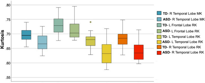
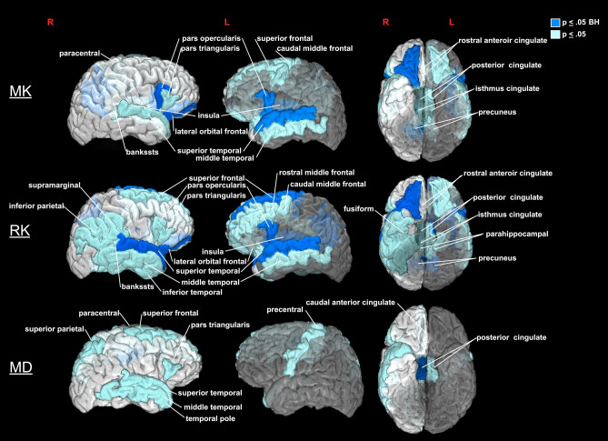
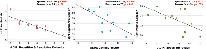
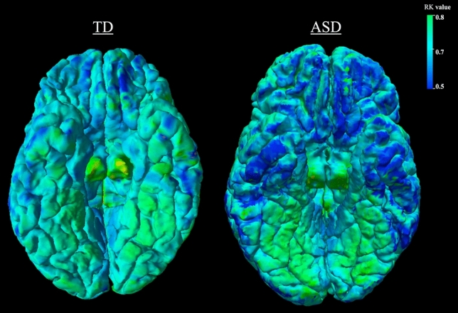
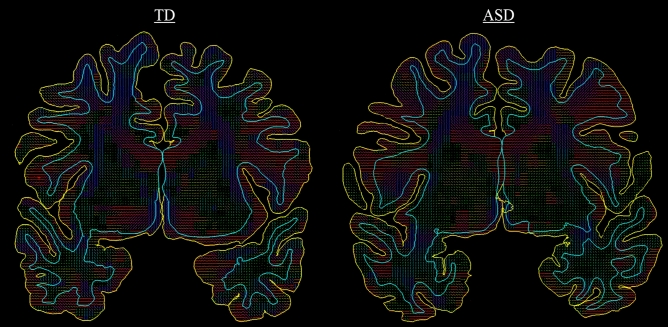
Prior ex vivo histological postmortem studies of autism spectrum disorder (ASD) have shown gray matter microstructural abnormalities, however, in vivo examination of gray matter microstructure in ASD has remained scarce due to the relative lack of non-invasive methods to assess it. The aim of this work was to evaluate the feasibility of employing diffusional kurtosis imaging (DKI) to describe gray matter abnormalities in ASD in vivo. DKI data were examined for 16 male participants with a diagnosis of ASD and IQ>80 and 17 age- and IQ-matched male typically developing (TD) young adults 18-25 years old. Mean (MK), axial (AK), radial (RK) kurtosis and mean diffusivity (MD) metrics were calculated for lobar and sub-lobar regions of interest. Significantly decreased MK, RK, and MD were found in ASD compared to TD participants in the frontal and temporal lobes and several sub-lobar regions previously associated with ASD pathology. In ASD participants, decreased kurtosis in gray matter ROIs correlated with increased repetitive and restricted behaviors and poor social interaction symptoms. Decreased kurtosis in ASD may reflect a pathology associated with a less restrictive microstructural environment such as decreased neuronal density and size, atypically sized cortical columns, or limited dendritic arborizations.
Conflict of interest statement
The authors declare no competing interests.
Figures
Similar articles
-
Diffusion kurtosis imaging biomarkers associated with amelioration of neuroinflammation, gray matter microstructural abnormalities, and gut dysbiosis by central thalamic deep brain stimulation in autistic -like young rats.Neuroimage. 2025 Aug 15;317:121344. doi: 10.1016/j.neuroimage.2025.121344. Epub 2025 Jun 21. Neuroimage. 2025. PMID: 40544899
-
Diffusion kurtosis imaging of gray matter in schizophrenia.Cortex. 2019 Dec;121:201-224. doi: 10.1016/j.cortex.2019.08.013. Epub 2019 Aug 29. Cortex. 2019. PMID: 31629198 Free PMC article.
-
In Vivo Evidence of Reduced Integrity of the Gray-White Matter Boundary in Autism Spectrum Disorder.Cereb Cortex. 2017 Feb 1;27(2):877-887. doi: 10.1093/cercor/bhw404. Cereb Cortex. 2017. PMID: 28057721 Free PMC article.
-
Diagnosing autism severity associated with physical fitness and gray matter volume in children with autism spectrum disorder: Explainable machine learning method.Complement Ther Clin Pract. 2024 Feb;54:101825. doi: 10.1016/j.ctcp.2023.101825. Epub 2023 Dec 30. Complement Ther Clin Pract. 2024. PMID: 38169278 Review.
-
Translational Neuroscience in Autism: From Neuropathology to Transcranial Magnetic Stimulation Therapies.Psychiatr Clin North Am. 2020 Jun;43(2):229-248. doi: 10.1016/j.psc.2020.02.004. Epub 2020 Apr 8. Psychiatr Clin North Am. 2020. PMID: 32439019 Free PMC article. Review.
Cited by
-
Automated predictive analytics tool for rainfall forecasting.Sci Rep. 2021 Sep 6;11(1):17704. doi: 10.1038/s41598-021-95735-8. Sci Rep. 2021. PMID: 34489507 Free PMC article.
-
Interplay of Neuroinflammation and Gut Microbiota Dysbiosis in Alzheimer's Disease Using Diffusion Kurtosis Imaging Biomarker in 3 × Tg-AD Mouse Models.ACS Chem Neurosci. 2025 Apr 16;16(8):1511-1528. doi: 10.1021/acschemneuro.5c00063. Epub 2025 Apr 7. ACS Chem Neurosci. 2025. PMID: 40195658 Free PMC article.
-
Application of Quantitative Magnetic Resonance Imaging in the Diagnosis of Autism in Children.Front Med (Lausanne). 2022 May 12;9:818404. doi: 10.3389/fmed.2022.818404. eCollection 2022. Front Med (Lausanne). 2022. PMID: 35646984 Free PMC article.
-
Reduced neurite density index in the prefrontal cortex of adults with autism assessed using neurite orientation dispersion and density imaging.Front Neurol. 2023 Aug 11;14:1110883. doi: 10.3389/fneur.2023.1110883. eCollection 2023. Front Neurol. 2023. PMID: 37638188 Free PMC article.
-
MR imaging and outcome in neonatal HIBD models are correlated with sex: the value of diffusion tensor MR imaging and diffusion kurtosis MR imaging.Front Neurosci. 2023 Sep 15;17:1234049. doi: 10.3389/fnins.2023.1234049. eCollection 2023. Front Neurosci. 2023. PMID: 37790588 Free PMC article.
References
-
- American Psychiatric Association. DSM-5 Diagnostic Classification. Diagnostic and Statistical Manual of Mental Disorders (5th ed.) (2013).
-
- Blumberg SJ, et al. Changes in prevalence of parent-reported autism spectrum disorder in school-aged U.S. children: 2007 to 2011–2012. Natl. Health Stat. Report. 2013;20:1–11. - PubMed
