Review
. 2021 Mar;17(3):150-161.
doi: 10.1038/s41574-020-00443-4. Epub 2020 Dec 8.

Type 1 diabetes mellitus as a disease of the β-cell (do not blame the immune system?)

Affiliations
Review

Type 1 diabetes mellitus as a disease of the β-cell (do not blame the immune system?)

Bart O Roep et al. Nat Rev Endocrinol. 2021 Mar.

Abstract

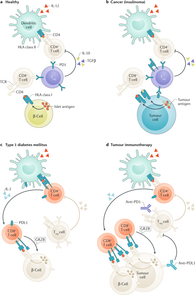
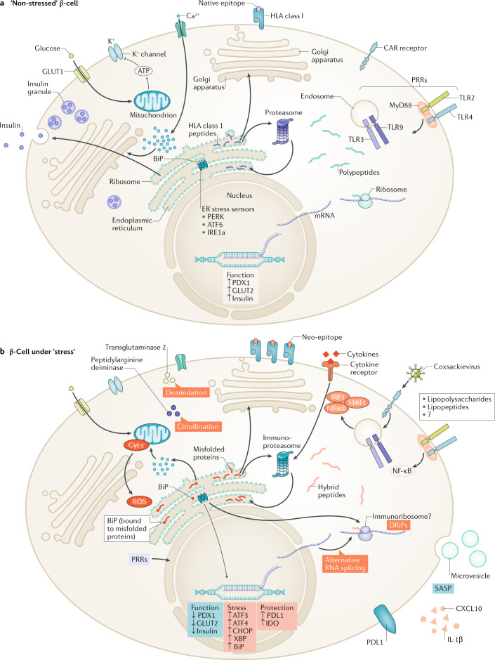
Type 1 diabetes mellitus is believed to result from destruction of the insulin-producing β-cells in pancreatic islets that is mediated by autoimmune mechanisms. The classic view is that autoreactive T cells mistakenly destroy healthy ('innocent') β-cells. We propose an alternative view in which the β-cell is the key contributor to the disease. By their nature and function, β-cells are prone to biosynthetic stress with limited measures for self-defence. β-Cell stress provokes an immune attack that has considerable negative effects on the source of a vital hormone. This view would explain why immunotherapy at best delays progression of type 1 diabetes mellitus and points to opportunities to use therapies that revitalize β-cells, in combination with immune intervention strategies, to reverse the disease. We present the case that dysfunction occurs in both the immune system and β-cells, which provokes further dysfunction, and present the evidence leading to the consensus that islet autoimmunity is an essential component in the pathogenesis of type 1 diabetes mellitus. Next, we build the case for the β-cell as the trigger of an autoimmune response, supported by analogies in cancer and antitumour immunity. Finally, we synthesize a model ('connecting the dots') in which both β-cell stress and islet autoimmunity can be harnessed as targets for intervention strategies.


Conflict of interest statement

The authors declare no competing interests.

Figures

Fig. 1
Fig. 1. Immunoregulation in health, and immune dysregulation in cancer, T1DM or immunotherapy.
a | In healthy individuals, β-cells are protected from autoimmune β-cell destruction by immune regulation exerted by regulatory T (Treg) cells and PD1 ligation. b | While advantageous in preventing autoimmunity, Treg cells impede antitumour immunity. c | In type 1 diabetes mellitus (T1DM), insufficient immune regulation can result in an autoimmune response by autoreactive T cells, particularly if these cells are provoked by β-cells. d | The response in T1DM resembles effective antitumour immunity as a result of immunotherapeutic blockade of PD1 or its ligand PDL1 that otherwise keep autoimmune responses in check. In addition to resulting in antitumour immunity, other immune and autoimmune responses might be triggered, including those against pancreatic islets. T1DM is a serious adverse effect of tumour immunotherapy. GRZB, granzyme B; TCR, T cell receptor.
Fig. 2
Fig. 2. The effect of stress on β-cell function and immunogenicity.
a | In non-stressed (‘happy’) β-cells, glucose uptake via the glucose transporter GLUT1 leads to pyruvate formation through glycolysis and to increased ATP production by mitochondria. The resulting increased cytosolic level of ATP promotes the closure of the potassium channels, membrane depolarization and opening of the voltage-dependent calcium channels. The rise in intracellular levels of calcium triggers exocytosis of the insulin-containing granules. In these conditions, several genes and proteins are upregulated to restore the cellular stock of insulin. During this process, non-translocated preproinsulin, endoplasmic reticulum (ER)-resident insulin signal peptide and non-mature proinsulin molecules are degraded through the proteasome directly or after retro-translocation and are presented by HLA class I at the cell surface (normal β-cell ligandome). b | In type 1 diabetes mellitus pathophysiological conditions, cytokines lead to profound changes in gene and protein expression, mainly by activation of the STAT1, IRF1 and NF-κB downstream pathways, that ultimately drives hyper-expression of HLA class I, and also the surface expression of inhibitory receptors (that is, PDL1). In the stress condition, calcium uptake by the mitochondria is responsible for increased permeability of the mitochondria that precedes the release of pro-apoptotic factors (such as reactive oxygen species (ROS) and cytochrome c (cyt c)). Calcium depletion in the ER leads to activation of cytosolic calcium-dependent enzymes (such as transglutaminase 2 and peptidyl arginine deiminase) that are involved in the post-translational modification processes by inducing protein deamidation and citrullination, respectively. The recruitment of the ER chaperones (binding immunoglobulin protein (BiP)) in response to the accumulation of misfolded protein within the ER leads to the activation of the sensors (PERK, IRE1a and ATF6) expressed at the surface of the ER membrane. The PERK pathway attenuates mRNA translation by phosphorylation of the eIF2a translation initiation factor. Phosphorylation of IRE1a and translocation of ATF6 activate the ER accumulated protein degradation pathway and the transcription of chaperone encoding genes with protein products involved in degradation of misfolded proteins, and also restores ER homeostasis. Long-term exposure to inflammatory stress promotes additional coping mechanisms, including initiation of recycling programmes and selective secretion of proteins and small RNAs in microvesicles, and ultimately leads to the induction of an apoptosis programme mediated by the transcriptional activation of CHOP by the combined activity of PERK and ATF6. During stress, normal transcription, translation and degradation is affected, which generates alternative RNA splicing, defective ribosomal products and hybrid peptides, respectively (stressed β-cell ligandome). CAR, coxsackievirus and adenovirus receptor; DRiP, defective ribosomal product; PRR, pattern recognition receptor; SASP, senescence-associated secretory phenotype; TLR, Toll-like receptor.

References

    1. Gepts W. Islet changes suggesting a possible immune aetiology of human diabetes mellitus. Acta Endocrinol. Suppl. 1976;205:95–106. - PubMed
    1. Bottazzo GF, et al. In situ characterization of autoimmune phenomena and expression of HLA molecules in the pancreas in diabetic insulitis. N. Engl. J. Med. 1985;313:353–360. doi: 10.1056/NEJM198508083130604. - DOI - PubMed
    1. Roep BO. The role of T-cells in the pathogenesis of type 1 diabetes: from cause to cure. Diabetologia. 2003;46:305–321. doi: 10.1007/s00125-003-1089-5. - DOI - PubMed
    1. Eizirik DL, Colli ML, Ortis F. The role of inflammation in insulitis and β-cell loss in type 1 diabetes. Nat. Rev. Endocrinol. 2009;5:219–226. doi: 10.1038/nrendo.2009.21. - DOI - PubMed
    1. Roep BO, Kracht MJ, van Lummel M, Zaldumbide A. A roadmap of the generation of neoantigens as targets of the immune system in type 1 diabetes. Curr. Opin. Immunol. 2016;43:67–73. doi: 10.1016/j.coi.2016.09.007. - DOI - PubMed

Publication types

MeSH terms