

MOB1A regulates glucose deprivation-induced autophagy via IL6-STAT3 pathway in gallbladder carcinoma
- PMID: 33294275
- PMCID: PMC7716168
MOB1A regulates glucose deprivation-induced autophagy via IL6-STAT3 pathway in gallbladder carcinoma
Abstract
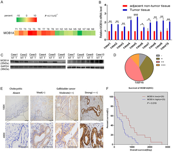
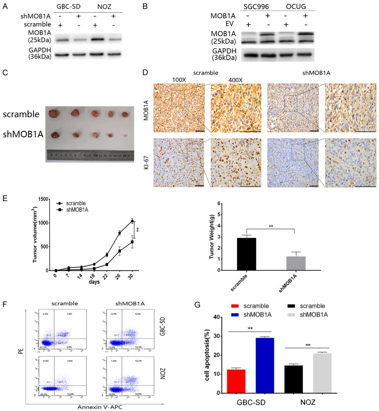
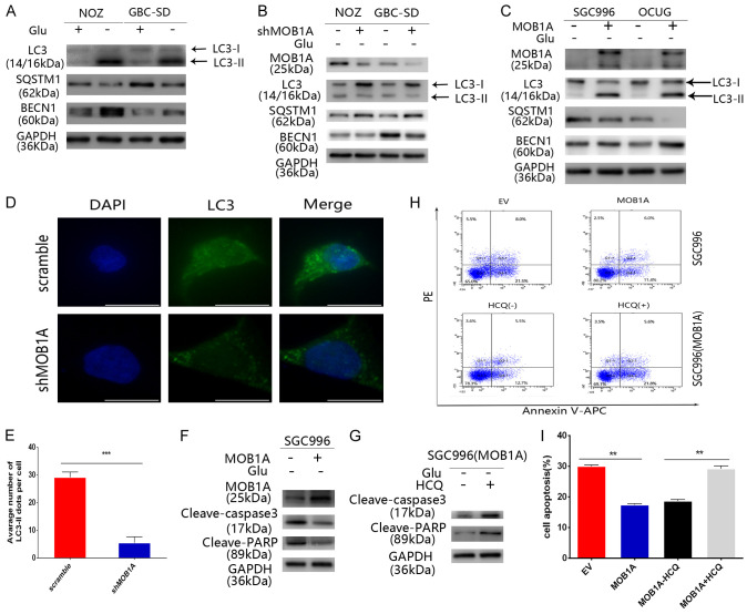
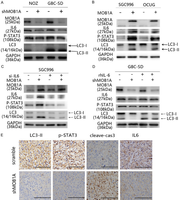
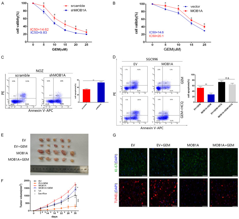
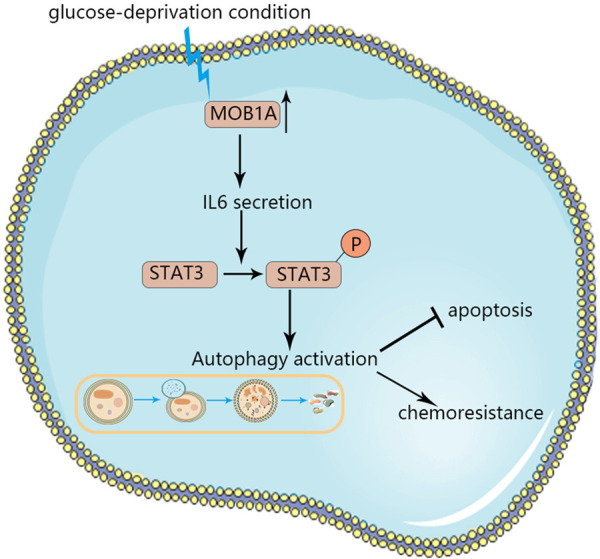
MOB kinase activator 1A (MOB1A) plays an important role in many diseases and cancers. Here, we observed that MOB1A was substantially overexpressed in gallbladder carcinoma (GBC) tissues compared with nontumor tissues. The high expression of MOB1A was closely associated with poor survival in patients with GBC at advanced TNM stages. Furthermore, our study indicated that MOB1A promoted autophagy by activating the IL6/STAT3 signaling pathway and regulating the chemosensitivity to gemcitabine under glucose deprivation conditions both in vitro and in vivo. In conclusion, these findings suggested that MOB1A is critical for the development of GBC via the MOB1A-IL6/STAT3-autophagy axis.
Keywords: IL6-STAT3 pathways; MOB1A; autophagy; gallbladder carcinoma; glucose-deprivation.
AJCR Copyright © 2020.
Conflict of interest statement
None.
Figures
Similar articles
-
MOB kinase activator 1A acts as an oncogene by targeting PI3K/AKT/mTOR in ovarian cancer.Discov Oncol. 2023 Jun 14;14(1):100. doi: 10.1007/s12672-023-00705-3. Discov Oncol. 2023. PMID: 37314589 Free PMC article.
-
Long noncoding RNA HEGBC promotes tumorigenesis and metastasis of gallbladder cancer via forming a positive feedback loop with IL-11/STAT3 signaling pathway.J Exp Clin Cancer Res. 2018 Aug 7;37(1):186. doi: 10.1186/s13046-018-0847-7. J Exp Clin Cancer Res. 2018. PMID: 30086773 Free PMC article.
-
Depletion of MOB1A/B causes intestinal epithelial degeneration by suppressing Wnt activity and activating BMP/TGF-β signaling.Cell Death Dis. 2018 Oct 22;9(11):1083. doi: 10.1038/s41419-018-1138-0. Cell Death Dis. 2018. PMID: 30349003 Free PMC article.
-
A novel tumor-promoting mechanism of IL6 and the therapeutic efficacy of tocilizumab: Hypoxia-induced IL6 is a potent autophagy initiator in glioblastoma via the p-STAT3-MIR155-3p-CREBRF pathway.Autophagy. 2016 Jul 2;12(7):1129-52. doi: 10.1080/15548627.2016.1178446. Epub 2016 May 10. Autophagy. 2016. PMID: 27163161 Free PMC article.
-
The role of STAT3 in autophagy.Autophagy. 2015;11(5):729-39. doi: 10.1080/15548627.2015.1017192. Autophagy. 2015. PMID: 25951043 Free PMC article. Review.
Cited by
-
Integrative analysis of exosomal ncRNAs and their regulatory networks in liver cancer progression.Pract Lab Med. 2025 Mar 21;45:e00464. doi: 10.1016/j.plabm.2025.e00464. eCollection 2025 Jul. Pract Lab Med. 2025. PMID: 40226122 Free PMC article.
-
Molecular mechanisms and therapeutic strategies in overcoming chemotherapy resistance in cancer.Mol Biomed. 2025 Jan 6;6(1):2. doi: 10.1186/s43556-024-00239-2. Mol Biomed. 2025. PMID: 39757310 Free PMC article. Review.
-
Boron Derivatives Inhibit the Proliferation of Breast Cancer Cells and Affect Tumor-Specific T Cell Activity In Vitro by Distinct Mechanisms.Biol Trace Elem Res. 2023 Dec;201(12):5692-5707. doi: 10.1007/s12011-023-03632-0. Epub 2023 Mar 20. Biol Trace Elem Res. 2023. PMID: 36940038
-
Up-regulation of MTHFD2 is associated with clinicopathological characteristics and poor survival in ovarian cancer, possibly by regulating MOB1A signaling.J Ovarian Res. 2022 Feb 8;15(1):23. doi: 10.1186/s13048-022-00954-w. J Ovarian Res. 2022. PMID: 35135596 Free PMC article.
-
MOB kinase activator 1A acts as an oncogene by targeting PI3K/AKT/mTOR in ovarian cancer.Discov Oncol. 2023 Jun 14;14(1):100. doi: 10.1007/s12672-023-00705-3. Discov Oncol. 2023. PMID: 37314589 Free PMC article.
References
-
- Li M, Liu F, Zhang F, Zhou W, Jiang X, Yang Y, Qu K, Wang Y, Ma Q, Wang T, Bai L, Wang Z, Song X, Zhu Y, Yuan R, Gao Y, Liu Y, Jin Y, Li H, Xiang S, Ye Y, Zhang Y, Jiang L, Hu Y, Hao Y, Lu W, Chen S, Gu J, Zhou J, Gong W, Zhang Y, Wang X, Liu X, Liu C, Liu H, Liu Y, Liu Y. Genomic ERBB2/ERBB3 mutations promote PD-L1-mediated immune escape in gallbladder cancer: a whole-exome sequencing analysis. Gut. 2019;68:1024–1033. - PubMed
-
- Xiang S, Wang Z, Ye Y, Zhang F, Li H, Yang Y, Miao H, Liang H, Zhang Y, Jiang L, Hu Y, Zheng L, Liu X, Liu Y. E2F1 and E2F7 differentially regulate KPNA2 to promote the development of gallbladder cancer. Oncogene. 2019;38:1269–1281. - PubMed
-
- Li M, Zhang Z, Li X, Ye J, Wu X, Tan Z, Liu C, Shen B, Wang XA, Wu W, Zhou D, Zhang D, Wang T, Liu B, Qu K, Ding Q, Weng H, Ding Q, Mu J, Shu Y, Bao R, Cao Y, Chen P, Liu T, Jiang L, Hu Y, Dong P, Gu J, Lu W, Shi W, Lu J, Gong W, Tang Z, Zhang Y, Wang X, Chin YE, Weng X, Zhang H, Tang W, Zheng Y, He L, Wang H, Liu Y, Liu Y. Whole-exome and targeted gene sequencing of gallbladder carcinoma identifies recurrent mutations in the ErbB pathway. Nat Genet. 2014;46:872–876. - PubMed
LinkOut - more resources
Full Text Sources
Miscellaneous