. 2020 Nov 25:2020:6644687.
doi: 10.1155/2020/6644687. eCollection 2020.

Effect of SIS3 on Extracellular Matrix Remodeling and Repair in a Lipopolysaccharide-Induced ARDS Rat Model

Affiliations

Effect of SIS3 on Extracellular Matrix Remodeling and Repair in a Lipopolysaccharide-Induced ARDS Rat Model

Qiong Liang et al. J Immunol Res. .

Abstract

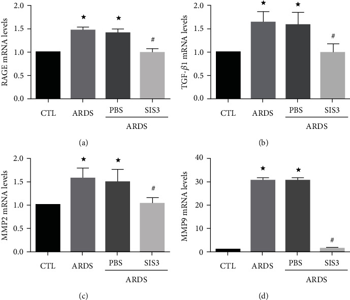
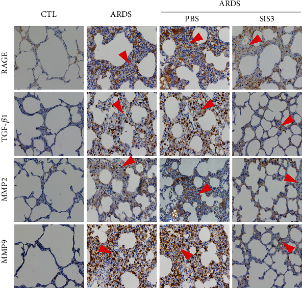
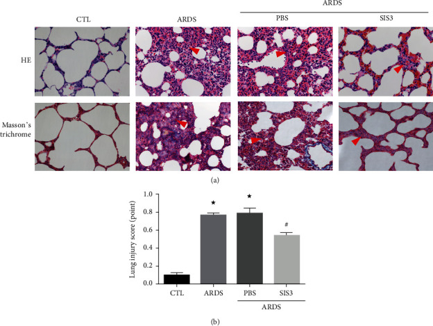
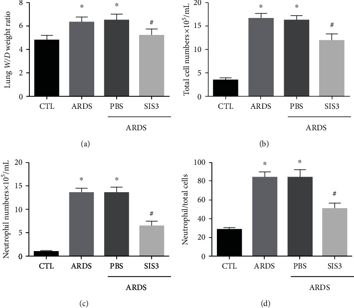
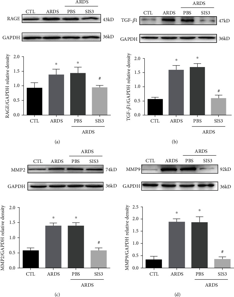
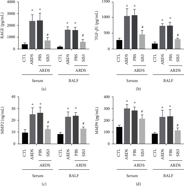
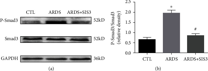
The remodeling of the extracellular matrix (ECM) in the parenchyma plays an important role in the development of acute respiratory distress syndrome (ARDS), a disease characterized by lung injury. Although it is clear that TGF-β1 can modulate the expression of the extracellular matrix (ECM) through intracellular signaling molecules such as Smad3, its role as a therapeutic target against ARDS remains unknown. In this study, a rat model was established to mimic ARDS via intratracheal instillation of lipopolysaccharide (LPS). A selective inhibitor of Smad3 (SIS3) was intraperitoneally injected into the disease model, while phosphate-buffered saline (PBS) was used in the control group. Animal tissues were then evaluated using histological analysis, immunohistochemistry, RT-qPCR, ELISA, and western blotting. LPS was found to stimulate the expression of RAGE, TGF-β1, MMP2, and MMP9 in the rat model. Moreover, treatment with SIS3 was observed to reverse the expression of these molecules. In addition, pretreatment with SIS3 was shown to partially inhibit the phosphorylation of Smad3 and alleviate symptoms including lung injury and pulmonary edema. These findings indicate that SIS3, or the blocking of TGF-β/Smad3 pathways, could influence remodeling of the ECM and this may serve as a therapeutic strategy against ARDS.


Conflict of interest statement

The authors declare that this research was conducted in the absence of any commercial or financial relationships that could be construed as a potential conflict of interest.

Figures

Figure 1
Figure 1
SIS3 inhibits mRNA expression of RAGE, TGF-β1, MMP2, and MMP9 in lung homogenates of ARDS rats. The mRNA levels of RAGE, TGF-β1, MMP2, and MMP9 were determined using real-time PCR and were standardized to β-actin. The results suggested that SIS3 inhibited the increase of (a) RAGE, (b) TGF-β1, (c) MMP2, and (d) MMP9 in LPS-induced lung homogenates of ARDS rats, while no effect was seen upon pretreatment with PBS. The data are presented as the means ± SD (n = 10). P < 0.05 vs. CTL; #P < 0.05 vs. ARDS.
Figure 2
Figure 2
SIS3 pretreatment decreases the protein expression and localization of RAGE, TGF-β1, MMP2, and MMP9 in ARDS rats. Immunohistochemical staining of lung tissue sections showed that RAGE, TGF-β1, MMP2, and MMP9 were expressed in the bronchial smooth muscle, airways, and alveolar epithelial cells of rats. The expression levels of RAGE, TGF-β1, MMP2, and MMP9 in the ECM of ARDS rats induced by LPS were significantly higher than those of the control group. RAGE, TGF-β1, MMP2, and MMP9 were reduced by pretreatment with SIS3, and no effect was found upon pretreatment with PBS. The micrographs were magnified at 400x. The red triangles indicate infiltrated leukocytes.
Figure 3
Figure 3
The inflammatory response was reduced by SIS3 pretreatment in LPS-induced acute lung injury. (a) Histological staining was performed to compare lung injury among the different groups. Both HE staining (upper panel) and Masson's trichrome staining (lower panel) show drastic leukocyte infiltration in the lung interstitium in the ARDS group, and the leukocyte infiltration was markedly alleviated after treatment of SIS3. Masson's trichrome staining reveals abnormal fibrosis and collagen distribution which was stained as blue, in the ARDS group, and significantly less blue stain is seen in the ARDS group treated with SIS3. The red triangles indicate the infiltrated sites. The photomicrographs were magnified to 400x. (b) HE staining was used to score lung injury. In the ARDS group, the lung injury was 0.77 ± 0.01, and after treatment with SIS3, the score decreased to 0.54 ± 0.01 (P < 0.05 vs. CTL; #P < 0.05 vs. ARDS). Data are presented as the means ± SD (n = 10).
Figure 4
Figure 4
SIS3 pretreatment alleviates lung injury and focal inflammation in LPS-induced ARDS. (a) BALF was collected 24 h after intratracheal instillation of LPS. The alternation of the alveolar capillary barrier was weakened by SIS3 pretreatment in ARDS rats, which was seen in an increase of the lung W/D ratio. (b–d) The total cells, neutrophils, and neutrophil ratio in the BALF were higher in the ARDS group than in the control group. The values were also lower in the ARDS + SIS3 group when compared to the ARDS group, respectively. The data are presented as the means ± SD (n = 10). P < 0.05 vs. CTL; #P < 0.05 vs. ARDS.
Figure 5
Figure 5
SIS3 inhibited the protein expression of RAGE, TGF-β1, MMP2, and MMP9 proteins in lung homogenates acquired from ARDS rats. The expression levels of RAGE, TGF-β1, MMP2, and MMP9 proteins in lung tissue homogenates, sera, and BALF of ARDS rats and were determined using western blotting analysis at 24 h after LPS intervention. The results of western blotting showed that SIS3 inhibited LPS-induced (a) RAGE, (b) TGF-β1, (c) MMP2, and (d) and MMP9 protein expression in the lung homogenates. The data are presented as the means ± SD (n = 10). P < 0.05 vs. CTL; #P < 0.05 vs. ARDS.
Figure 6
Figure 6
SIS3 pretreatment prevented the reduction of the expression of RAGE, TGF-β1, MMP2, and MMP9 in LPS-induced ARDS. The effects of SIS3 on the protein expression levels of (a) RAGE, (b) TGF-β1, (c) MMP2, and (d) MMP9 in the BALF and sera of the ARDS rats were determined using ELISA. The results demonstrated that the levels of RAGE, TGF-β1, MMP2, and MMP9 proteins were significantly higher in the ARDS group than in the control group, while the levels of RAGE, TGF-β1, MMP2, and MMP9 in the SIS3 group were lower than those in the ARDS group. The protein levels of RAGE, TGF-β1, MMP2, and MMP9 in LPS-administered pretreatment with PBS were not different from those of the ARDS group. The data are presented as the means ± SD of three independent experiments in triplicate (n = 10). P < 0.05 vs. CTL; #P < 0.05 vs. ARDS.
Figure 7
Figure 7
Phosphorylation of smad3 was inhibited by SIS3 in the lung tissue of ARDS rats. The activation of Smad3 was evaluated using western blotting analysis, and the amount of phospho-Smad3 formed was analyzed 24 h after treatment with either LPS or PBS. GAPDH was used as the loading control. The data are presented as the means ± SD (n = 10). P < 0.05 vs. CTL; #P < 0.05 vs. ARDS.

References

    1. Adachi N., Numakawa T., Richards M., Nakajima S., Kunugi H. New insight in expression, transport, and secretion of brain-derived neurotrophic factor: implications in brain-related diseases. World Journal of Biological Chemistry. 2014;5(4):409–428. doi: 10.4331/wjbc.v5.i4.409. - DOI - PMC - PubMed
    1. Azoulay E., Citerio G., Bakker J., et al. Year in review in Intensive Care Medicine 2013: II. Sedation, invasive and noninvasive ventilation, airways, ARDS, ECMO, family satisfaction, end-of-life care, organ donation, informed consent, safety, hematological issues in critically ill patients. Intensive Care Medicine. 2014;40(3):305–319. doi: 10.1007/s00134-014-3217-8. - DOI - PubMed
    1. Aschner Y., Zemans R. L., Yamashita C. M., Downey G. P. Matrix metalloproteinases and protein tyrosine kinases. Chest. 2014;146(4):1081–1091. doi: 10.1378/chest.14-0397. - DOI - PMC - PubMed
    1. Hergrueter A. H., Nguyen K., Owen C. A. Matrix metalloproteinases: all the RAGE in the acute respiratory distress syndrome. American Journal of Physiology. Lung Cellular and Molecular Physiology. 2011;300(4):L512–L515. doi: 10.1152/ajplung.00023.2011. - DOI - PMC - PubMed
    1. Sharp C., Millar A. B., Medford A. R. L. Advances in understanding of the pathogenesis of acute respiratory distress syndrome. Respiration. 2015;89(5):420–434. doi: 10.1159/000381102. - DOI - PubMed

MeSH terms

Substances

LinkOut - more resources