. 2020 Oct 27:9:310-318.
doi: 10.1016/j.ibror.2020.10.005. eCollection 2020 Dec.

Ventral root evoked entrainment of disinhibited bursts across early postnatal development in mice

Affiliations

Ventral root evoked entrainment of disinhibited bursts across early postnatal development in mice

Chetan Nagaraja. IBRO Rep. .

Abstract

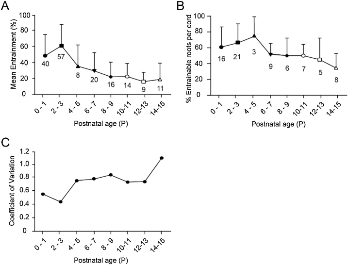
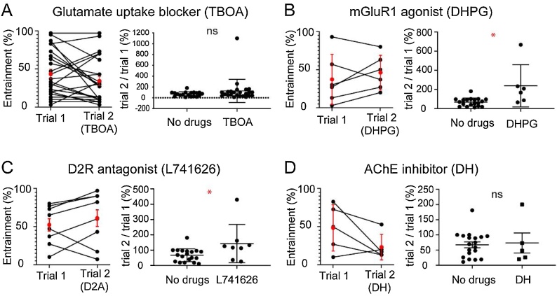
Early in the postnatal period, motoneuron axon stimulation can excite motor networks in the spinal cord. Here we tested if these excitatory effects changed across early postnatal development up to postnatal day (P) 24 by when mice are capable of weight-bearing locomotion and locomotor networks are considered functionally mature. This was accomplished in the isolated spinal cord preparation using ventral root evoked entrainment of disinhibited bursts. Ventral root evoked entrainment was defined and characterized over the first 2 weeks of postnatal development, and was found to decline over this period, but entrainment could still be detected in mice as old as P24. Disinhibited bursting could be elicited, and dorsal root evoked entrainment could be recorded as late as P39 and remained unchanged in effectiveness, suggesting that poor tissue viability may not be the cause of the decline in ventral root evoked entrainment. Pharmacological experiments performed on younger animals established that dopamine D2 receptor antagonists and mGluR1 agonists both enhanced ventral root evoked entrainment. In conclusion, the motoneuronal inputs to spinal motor networks via the excitatory pathway is modulated by dopamine and metabotropic glutamate receptors and may be under powerful inhibitory control, which may explain why there is a developmental decline in entrainment.

Keywords: D2, dopamine receptor subtype 2; DH, donepezil hydrochloride; DHPG, (RS)-3,5-dihydroxyphenylglycine; Disinhibited bursting; Entrainment; LLA, locomotor-like activity; Motoneurons; Network function; P, postnatal day; Spinal cord; TBOA, DL-Threo-β-Benzyloxyaspartic acid; mGluR1, metabotropic glutamate receptor subtype 1.


Figures

Fig. 1
Fig. 1
Characteristics of disinhibited bursting and entrainment by ventral root stimulations. A. Schematic of the spinal cord preparations used. B. The period of the disinhibited bursting is plotted for P0 – P15. The definition of the period is shown over the plot. Numbers on the plot indicate the number of intact or hemicord preparations used at each age. C. An example of a series of stimulus trains applied to the ventral root of a P0 cord to entrain disinhibited bursting. Filled triangles below the trace indicate instances of entrainment. Open triangles indicate stimulus trains that did not produce entrainment. Arrows indicate entrained bursts at a faster timescale (lower panel). D. Distribution of latencies from stimulus for a sequence of stimulus trains applied to entrain disinhibited bursting. Data from 22 ventral roots in 15 mice, ages P0 – P3. The inset plot shows the distribution of latencies from 0 to 3 seconds at enlarged timescale. E. A short segment of disinhibited bursting record from a P3 cord showing when the assumed stimuli (dashed lines) would have been applied. F. Distribution of latency (time from the dotted line to the next burst) for the record in panel E. 5.8 % of the latencies occurred from 0 to 1.5 s for the inter-train interval of 40 s.
Fig. 2
Fig. 2
Characterization of ventral root evoked entrainment from P0 to P15. A. Changes in mean value of entrainment at the different ages. Numbers on the plot indicate the number of ventral roots used at each age. B. Changes in percentage of entrainable roots per cord at the different ages. Numbers on the plot indicate the number of animals used at each age. C. Change in the coefficient of variation for the entrainment data at the different ages. Error bars are SD. P0-P9 are intact cord preparations. P10-15 are hemicord preparations.
Fig. 3
Fig. 3
Entrainment of disinhibited bursting at the oldest ages. A. Example of dorsal root evoked entrainment in a P38 hemicord. Arrowheads indicate the sequence of single stimulus (5 μA) applied to the L5 dorsal root. B. Example of ventral root evoked entrainment in a P24 hemicord. Arrowheads indicate a sequence of stimulus trains applied to the L5 ventral root. A and B. Filled arrowheads below the traces indicate instances of entrainment. Open arrowheads indicate that there was no entrainment. Arrows indicate entrained bursts at a faster timescale (lower panel).
Fig. 4
Fig. 4
Ventral root evoked entrainment decays following a 30-stimulus trial and partly recovers. A. Experimental protocol to assess time-dependent changes in entrainment. B. Plots showing change in entrainment for each experiment. Entrainment values for the two trials for each root are joined by lines, average of entrainment values for each trial are indicated by red filled circles and error bars indicate the standard deviation. Data for short inter-trial interval includes 9 ventral roots from 6 animals aged P0-P5, and that for long inter-trial intervals include 19 ventral roots from 14 animals aged P0-P4. C. Plot showing normalized change of entrainment for the experiments with short and long inter-trial intervals.
Fig. 5
Fig. 5
The effects of various drugs on entrainment. A – F. The entrainment values for trials 1 (before drugs) and 2 (after addition of drugs) are shown in the graphs on the left in each panel. The red filled circles indicate the mean and error bars indicate the standard deviation. The normalized change of entrainment (trial 2/ trial 1; mean ± SD) for the experiments with and without drugs (No drugs) are shown in the graphs on the right in each panel. * p < 0.5 **** p < 0.005. See text for further details.

Similar articles

Cited by

References

    1. Alvarez F.J., Fyffe R.E.W. The continuing case for the Renshaw cell. J. Physiol. 2007;584(1):31–45. - PMC - PubMed
    1. Bhumbra G.S., Beato M. Recurrent excitation between motoneurones propagates across segments and is purely glutamatergic. PLoS Biol. 2018;16(3) - PMC - PubMed
    1. Bhumbra G.S., Bannatyne B.A., Watanabe M., Todd A.J., Maxwell D.J., Beato M. The recurrent case for the renshaw cell. J. Neurosci. 2014;34(38):12919–12932. - PMC - PubMed
    1. Biscoe T.J., Nickels S.M., Stirling C.A. Numbers and sizes of nerve fibres in mouse spinal roots. Q. J. Exp. Physiol. 1982;67(3):473–494. - PubMed
    1. Bonnot A., Chub N., Pujala A., O’Donovan M.J. Excitatory actions of ventral root stimulation during network activity generated by the disinhibited neonatal mouse spinal cord. J. Neurophysiol. 2009;101(6):2995–3011. - PMC - PubMed

LinkOut - more resources