. 2021 Jan;25(2):1140-1150.
doi: 10.1111/jcmm.16180. Epub 2020 Dec 9.

Ubiquitously specific protease 4 inhibitor-Vialinin A attenuates inflammation and fibrosis in S100-induced hepatitis mice through Rheb/mTOR signalling

Affiliations

Ubiquitously specific protease 4 inhibitor-Vialinin A attenuates inflammation and fibrosis in S100-induced hepatitis mice through Rheb/mTOR signalling

Jie Xu et al. J Cell Mol Med. 2021 Jan.

Abstract

Inflammation and fibrosis are major consequences of autoimmune hepatitis, however, the therapeutic mechanism remains to be investigated. USP4 is a deubiquitinating enzyme and plays an important role in tissue fibrosis and immune disease. Vialinin A is an extract from mushroom and is a specific USP4 inhibitor. However, there is lack of evidences that Vialinin A plays a role in autoimmune hepatitis. By employing S100-induced autoimmune hepatitis in mice and AML12 cell line, therapeutic mechanism of Vialinin A was examined. Inflammation was documented by liver histological staining and inflammatory cytokines. Fibrosis was demonstrated by Masson, Sirius red staining and fibrotic cytokines with western blot and real-time RT-PCR. In experimental animal, there were increases in inflammation and fibrosis as well as USP4, and which were reduced after treatment of Vialinin A. Vialinin A also reduced Rheb and phosphorylated mTOR. Moreover, in LPS-treated AML12 cells, LPS-induced USP4, inflammatory and fibrotic cytokines, phosphorylated mTOR and Rheb. Specific inhibitory siRNA of USP4 reduced USP4 level and the parameters mentioned above. In conclusion, USP4 was significantly elevated in autoimmune hepatitis mice and Vialinin A reduced USP4 level and attenuate inflammation and fibrosis in the liver. The mechanism may be related to regulation of Rheb/mTOR signalling.

Keywords: USP4; Vialinin A; autoimmune hepatitis; mTOR.


Conflict of interest statement

All authors have no potential conflict of interest to declare. All authors read and approved of the final version of the manuscript. All authors have read the journal's policy on conflicts of interest. All authors have read the journal's authorship agreement.

Figures

FIGURE 1
FIGURE 1
Vialinin A attenuates the inflammation and fibrosis of the liver in AIH mice. Panel A shows the protocol of establishment of autoimmune hepatitis in mice and Via injection. Panel B displays the serum levels of ALT and AST from each group, IgG from Ctrl and AIH group. Panel C shows the representative pictures of H&E, Masson and Sirius red staining of liver tissues. Black triangle highlights the lymphocytic infiltration (magnification, ×200). The right panels represent quantification of the fibrotic area from Masson staining and Sirius red staining. The data are presented as mean ± SED from 6 mice. ***P < .001
FIGURE 2
FIGURE 2
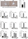
The expression of USP4 and the mRNA levels of inflammatory cytokines in the liver of AIH mice and AML12 cells. Panel A shows representative immunohistochemistry staining (magnification, ×400) of USP4 and the USP4 expression score in liver of normal mice and AIH model. Panel B displays the histogram of relative mRNA levels of USP4, IL‐1 and IL‐6 in the liver from each group. Panel C shows the histogram of relative mRNA level from AML12 cells treated with LPS plus or minus Vialinin A or specific inhibitory siRNA of UPS4. GAPDH used as loading control. The data are presented as mean ± SDM from 6 mice or 3 independent experiments of cells. **P < .01 and ***P < .001
FIGURE 3
FIGURE 3
The protein levels of USP4 and fibrotic cytokines in mice and AML12 cells. Panel A shows the representative Western blot and histogram of proteins for USP4, COL‐1, α‐SMA, TGF‐β and FN14 in from each group. Panel B displays the typical Western blot and histogram of proteins of fibrotic cytokines from AML12 cells treated with LPS plus or minus Vialinin A. Panel C shows the typical Western blot and histogram of proteins different fibrotic cytokines proteins from AML12 cells treated with LPS specific inhibitory siRNA of UPS4. GAPDH used as loading control. The data represents mean ± SDM from 6 mice or 3 independent experiments from cells. *P < .05, **P < .01 and ***P < .001
FIGURE 4
FIGURE 4
The mRNA levels of fibrosis markers from experimental mice and AML12 cells. Panel A displays relative mRNA levels of COL‐1, α‐SMA, TGF‐β and FN14 from each group. Panel B shows the relative mRNA levels of fibrosis markers from AML12 cells treated with LPS plus or minus Vialinin A. Panel C displays the same mRNA levels from AML12 cells treated with LPS alone or LPS with non‐specific or specific inhibitory siRNA of USP4. GAPDH used as loading control. The data are presented as mean ± SDM. *P < .05, **P < .01 and ***P < .001
FIGURE 5
FIGURE 5
Alternation of Rheb/mTOR signalling in experimental mice and AML12 cells. Panel A shows representative Western blot pictures on the right and histigram of the band density on the right of Rheb, p‐mTOR, mTOR in the liver of each experimental group while panel B shows the same from AML12 cells treated with LPS plus or minus Vialinin A and panel C display the same form AML12 cells treated with LPS alone or LPS with non‐specific and specific inhibitory siRNA of UPS4. GAPDH used as loading control. The middle column shows the ratio of phosphorylated mTOR to mTOR. The data are presented as mean ± SDM. *P < .05, **P < .01 and ***P < .001
FIGURE 6
FIGURE 6
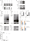
Immunoprecipitation experiments shows USP4 and Vialinini A involved in regulation of Rheb deubiquitination. Panel A shows the Rheb protein level in AML12 cells treated with 5 µmol/L LPS for 24 h and further treated with different protein inhibitors individually or in combination for 6 h of CHX (100 mg/mL), lysosomal inhibitor chloroquine (50 µmol/L) with and proteasome inhibitor MG132 (5 µmol/L). Panel B shows the immunoprecipated with USP4 and immunoblot with USP4 and Rheb with immunoprecipitaed proteins. Panel C displays the immunoprecipation experiments with proteasome inhibitor MG132 and LPS as well as with and without specific inhibitory siRNA of USP4. Protein was immunoprecipitated with antibody of Rheb and immunoblotted with Ubiquitin antibody. Panel d shows the same immunoprecipitation experiments as in panel C except with Vialinin A instead of specific inhibitory siRNA of USP4. The right Panel displays the histogram of (C) and (D). Panel e shows USP4 mRNA level from healthy and AIH patients. The data are presented as mean ± SDM from 6 mice of 3 independent experiments of cells. **P < .01 and ***P < .001

References

    1. Webb GJ, Hirschfield GM, Krawitt EL, Gershwin ME. Cellular and molecular mechanisms of autoimmune hepatitis. Annu Rev Pathol. 2018;13:247‐292. - PubMed
    1. Lapierre P, Béland K, Alvarez F. Pathogenesis of autoimmune hepatitis: from break of tolerance to immune‐mediated hepatocyte apoptosis. Transl Res. 2007;149(3):107‐113. - PubMed
    1. European Association for the Study of the Liver . EASL clinical practice guidelines: autoimmune hepatitis. J Hepatol. 2015;63(4):971‐1004. - PubMed
    1. Dyson JK, De Martin E, Dalekos GN, et al. Review article: unanswered clinical and research questions in autoimmune hepatitis‐conclusions of the International Autoimmune Hepatitis Group Research Workshop. Aliment Pharmacol Ther. 2019;49(5):528–536. - PubMed
    1. Trivedi PJ, Hubscher SG, Heneghan M, Gleeson D, Hirschfield GM. Grand round: autoimmune hepatitis. J Hepatol. 2019;70(4):773–784. - PubMed

Publication types

MeSH terms