Crocin inhibits the activation of mouse hepatic stellate cells via the lnc-LFAR1/MTF-1/GDNF pathway
- PMID: 33295246
- PMCID: PMC7781632
- DOI: 10.1080/15384101.2020.1848064
Crocin inhibits the activation of mouse hepatic stellate cells via the lnc-LFAR1/MTF-1/GDNF pathway
Abstract
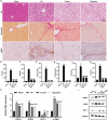
Crocin is the main monomer of saffron, which is a momentous component of traditional Chinese medicine Lang Qing A Ta. Here, we tried to probe into the role of crocin in liver fibrosis. Hematoxylin-eosin staining and Sirius Red staining were used to observe the pathological changes of liver tissues. After hepatic stellate cells (HSCs) were isolated from liver tissues, lnc-LFAR1, MTF-1, GDNF, and α-SMA expressions were detected by qRT-PCR and western blot. Immunohistochemistry and immunofluorescence were used to detect α-SMA expression. Chromatin immunoprecipitation was used to analyze the binding of MTF-1 to the GDNF promoter. Moreover, the dual-luciferase reporter gene, RNA pull-down, and RNA immunoprecipitation were used to clarify the interaction between MTF-1 and GDNF, lnc-LFAR1 and MTF-1. The degree of liver fibrosis was more severe in the mice from the liver fibrosis model, while the liver fibrosis was alleviated by the injection of crocin. lnc-LFAR1, GDNF, and α-SMA were up-regulated, and MTF-1 was down-regulated in liver fibrosis tissues and cells, while these trends were reversed after the injection of crocin. Besides, lnc-LFAR1 negatively regulated MTF-1 expression, and positively regulated GDNF and α-SMA expressions, and MTF-1 was enriched in the promoter region of GDNF. Furthermore, the cellular direct interactions between MTF-1 and GDNF, lnc-LFAR1 and MTF-1 were verified. In vivo experiments confirmed the relief of crocin on liver fibrosis. Our research expounded that crocin restrained the activation of HSCs through the lnc-LFAR1/MTF-1/GDNF axis, thereby ameliorating liver fibrosis.
Keywords: Crocin; GDNF; MTF-1; hepatic stellate cells; lnc-LFAR1.
Conflict of interest statement
No potential conflict of interest was reported by the authors.
Figures


Similar articles
-
The liver-enriched lnc-LFAR1 promotes liver fibrosis by activating TGFβ and Notch pathways.Nat Commun. 2017 Jul 26;8(1):144. doi: 10.1038/s41467-017-00204-4. Nat Commun. 2017. PMID: 28747678 Free PMC article.
-
Huangqi Decoction, a compound Chinese herbal medicine, inhibits the proliferation and activation of hepatic stellate cells by regulating the long noncoding RNA-C18orf26-1/microRNA-663a/transforming growth factor-β axis.J Integr Med. 2023 Jan;21(1):47-61. doi: 10.1016/j.joim.2022.11.002. Epub 2022 Nov 16. J Integr Med. 2023. PMID: 36456413
-
Glial cell line-derived neurotrophic factor (GDNF) mediates hepatic stellate cell activation via ALK5/Smad signalling.Gut. 2019 Dec;68(12):2214-2227. doi: 10.1136/gutjnl-2018-317872. Epub 2019 Jun 6. Gut. 2019. PMID: 31171625 Free PMC article.
-
Silencing lncRNA Lfar1 alleviates the classical activation and pyoptosis of macrophage in hepatic fibrosis.Cell Death Dis. 2020 Feb 18;11(2):132. doi: 10.1038/s41419-020-2323-5. Cell Death Dis. 2020. PMID: 32071306 Free PMC article.
-
c-Myc affects hedgehog pathway via KCNQ1OT1/RAC1: A new mechanism for regulating HSC proliferation and epithelial-mesenchymal transition.Dig Liver Dis. 2021 Nov;53(11):1458-1467. doi: 10.1016/j.dld.2020.11.035. Epub 2021 Jan 13. Dig Liver Dis. 2021. PMID: 33451909
Cited by
-
Noncoding RNA-Mediated Epigenetic Regulation in Hepatic Stellate Cells of Liver Fibrosis.Noncoding RNA. 2024 Aug 7;10(4):44. doi: 10.3390/ncrna10040044. Noncoding RNA. 2024. PMID: 39195573 Free PMC article. Review.
-
Sarcoma protein kinase inhibition alleviates liver fibrosis by promoting hepatic stellate cells ferroptosis.Open Life Sci. 2023 Dec 6;18(1):20220781. doi: 10.1515/biol-2022-0781. eCollection 2023. Open Life Sci. 2023. PMID: 38077794 Free PMC article.
-
Quercetagetin alleviates liver fibrosis in non-alcoholic fatty liver disease by promoting ferroptosis of hepatic stellate cells through GPX4 ubiquitination.Chin Med. 2025 Jun 16;20(1):89. doi: 10.1186/s13020-025-01109-x. Chin Med. 2025. PMID: 40524220 Free PMC article.
-
Molecular typing and prognostic model of lung adenocarcinoma based on cuprotosis-related lncRNAs.J Thorac Dis. 2022 Dec;14(12):4828-4845. doi: 10.21037/jtd-22-1534. J Thorac Dis. 2022. PMID: 36647499 Free PMC article.
-
Crocin's role in modulating MMP2/TIMP1 and mitigating hypoxia-induced pulmonary hypertension in mice.Sci Rep. 2024 Jun 3;14(1):12716. doi: 10.1038/s41598-024-62900-8. Sci Rep. 2024. PMID: 38830933 Free PMC article.
References
Publication types
MeSH terms
Substances
LinkOut - more resources
Full Text Sources
Other Literature Sources
Medical
Miscellaneous
