Review
. 2022 Jan;55(1):81-99.
doi: 10.1002/jmri.27411. Epub 2020 Dec 9.

Real-Time Magnetic Resonance Imaging

Affiliations
Review

Real-Time Magnetic Resonance Imaging

Krishna S Nayak et al. J Magn Reson Imaging. 2022 Jan.

Abstract

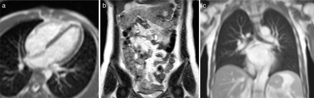
Real-time magnetic resonance imaging (RT-MRI) allows for imaging dynamic processes as they occur, without relying on any repetition or synchronization. This is made possible by modern MRI technology such as fast-switching gradients and parallel imaging. It is compatible with many (but not all) MRI sequences, including spoiled gradient echo, balanced steady-state free precession, and single-shot rapid acquisition with relaxation enhancement. RT-MRI has earned an important role in both diagnostic imaging and image guidance of invasive procedures. Its unique diagnostic value is prominent in areas of the body that undergo substantial and often irregular motion, such as the heart, gastrointestinal system, upper airway vocal tract, and joints. Its value in interventional procedure guidance is prominent for procedures that require multiple forms of soft-tissue contrast, as well as flow information. In this review, we discuss the history of RT-MRI, fundamental tradeoffs, enabling technology, established applications, and current trends. LEVEL OF EVIDENCE: 5 TECHNICAL EFFICACY STAGE: 1.

Keywords: fast imaging; interactive imaging; real-time MRI.


Figures

FIGURE 1:
FIGURE 1:
Publications involving RT-MRI. PubMed search: ((“real-time MRI”) OR (“real-time NMR”) OR (“real-time magnetic resonance”) OR (“real-time interactive MRI”) OR (“RT-MRI”)).
FIGURE 2:
FIGURE 2:
Scatterplot of 2D RT-MRI spatial and temporal resolution. Spatial resolution (x-axis) vs. temporal resolution (y-axis) is plotted from 22 recent publications that utilize state-of-the-art methodology, as selected by the authors of this review, summarized in Table S1. The gray shaded bar indicates the general spatiotemporal resolution tradeoff. All substantial deviations are due to variations in the FOV, use of parallel imaging, use of reconstruction constraints, and minimum acceptable SNR.
FIGURE 3:
FIGURE 3:
Common sequences, sampling trajectories, and view orders used in 2D RT-MRI. (a) Sequence diagrams of spoiled GRE and bSSFP, and the steady-state signal amplitude as a function of off-resonance Δf; Simulation parameters: TR = 5 msec; flip angle = 5° for spoiled GRE; flip angle = 60° for bSSFP; myocardium T1/T2 = 950/50 msec; blood T1/T2 = 1500/250 msec (representative of 1.5T). (b) Non-Cartesian sampling trajectories of undersampled radial, single-shot spiral, and single-shot EPI. (c) View orders of multishot spiral of conventional 13-interleaf bit-reversed and golden-ratio, and unaliased FOV as a function of the number of interleaves [Reproduced from Ref. (13)].
FIGURE 4:
FIGURE 4:
Illustration of cardiovascular RT-MRI. (a) Real-time cine imaging using tiny-golden angle radial bSSFP sequence at 1.5T, with 12x undersampling and compressive sensing reconstruction (TE/TR = 1.3/2.7 msec, flip angle = 70°, in-plane resolution = 2.1 mm, 32 msec temporal resolution, 31 fps). A movie can be found in Movie S1 [Adapted from Ref. (91)]. (b) Real-time PCMR using perturbed spirals at 1.5T, with 18x undersampling and compressive sensing reconstruction. Top: Magnitude images, Bottom: Phase images (TE/TR = 1.9/6.7 msec, VENC = 200 cm/s, flip angle = 20°, in-plane resolution = 1.8 mm, 27 msec temporal resolution, 37 fps). A movie can be found in Movie S2 [Adapted from Ref. (94)]. (c) Real-time imaging of the fetal heart (shown by arrow in first column) demonstrating gross fetal movement. Golden-angle radial bSSFP sequence at 1.5T, with 27x undersampling and compressive sensing reconstruction (TR = 5.0 msec, flip angle = 70°, in-plane resolution = 1.0 mm, 74 msec temporal resolution, 14 fps). A movie can be found in Movie S3 [Adapted from Ref. (95)].
FIGURE 5:
FIGURE 5:
Illustrations of RT-MRI for MRI-guided invasive procedures. Cardiovascular procedures are the most technically demanding for RT-MRI, and therefore are provided. (a) The position and orientation of catheter devices with two embedded microcoils are tracked on a previously acquired 3D volume for an electrophysiology procedure. Real-time device tracing is achieved using 3D gradient echo projection imaging (resolution 0.83 mm, 10 Hz tracking rate) [Reproduced from Ref. (114)]. (b) Interactive RT-MRI used to navigate gadolinium-filled balloon wedge end-hole catheter during diagnostic right heart catheterization (bSSFP, TE/TR = 1.44/2.88 msec, flip angle = 40°, in-plane resolution = 1.8×2.4 mm2, GRAPPA rate 2, 200 msec temporal resolution, 5 fps) [Adapted from Ref. (115)]. (c) Real-time MRI thermometry used to calculate thermal dose during therapeutic ablation procedure (gradient echo EPI, TE/TR = 18–20/110 msec, flip angle = 60, in-plane resolution = 1.6×1.6 mm2, GRAPPA rate 2, 200 msec temporal resolution, 5 slices/s) [Adapted from Ref. (116)].
FIGURE 6:
FIGURE 6:
Illustration of upper airway RT-MRI. (a) Speech production imaging using 13-interleave spiral GRE sequence at 1.5T (TE/TR = 0.8/6.0 msec, flip angle = 15° in-plane resolution = 2.4 mm, 12 msec temporal resolution, 83 fps) [Adapted from Ref. (142)]. (b) Sleep apnea study using simultaneous multislice radial GRE sequence at 3T (TE/TR = 3.7/6.5 msec, flip angle = 5°, slice thickness/gap = 7/3 mm, 3 slices, in-plane resolution = 1 mm, 96 msec temporal resolution, 10 fps) [Adapted from Ref. (37)]. (c) Swallowing imaging of 10-ml pineapple juice using radial FLASH sequence (TE/TR = 1.33/2.10 msec, flip angle = 8°, in-plane resolution = 1.3 mm, 40 msec temporal resolution, 25 fps, 19 spokes) [Adapted from Ref. (143)].
FIGURE 7:
FIGURE 7:
Illustration of three musculoskeletal RT-MRI applications. (a) Knee RT-MRI highlighting flexion, extension, and the measurement of rectus femoris knee muscle moment arms [Adapted from Ref. (164)]. (b) Wrist RT-MRI illustrating a radial–ulnar deviation maneuver, suitable for measuring dynamics of the scapholunate gap [Adapted from Ref. (165)]. (c) Temporomandibular joint RT-MRI illustrating the ability to track condyle movement during voluntary opening of the mouth [Adapted from Ref. (166)].
FIGURE 8:
FIGURE 8:
Illustration of three ML/AI-based low-latency applications. (a) Image reconstruction of cardiovascular imaging; (left-to-right) the BH-bSSFP sequence and the RT radial sequence reconstructed with gridding, GRASP, and the residual U-Net [Adapted from Ref. (93)] (b) Spiral off-resonance deblurring of speech imaging; (left-to-right) GT, uncorrected, IR with GT field map, and the CNN [Adapted from Ref. (78)]. (C) Needle detection and segmentation for ex vivo tissue RT-MRI; (left-to-right) Original image, needle detection and segmentation result using Mask R-CNN, result comparison against a reference [Adapted from Ref. (197)]. Note that “processing time” shown here is the time to run the neural networks and does not include the time to do preprocessing of the data. BH: breath-hold, GRASP: Golden-angle radial sparse parallel imaging, PT: processing time, GT: ground truth, IR: iterative reconstruction.
FIGURE 9:
FIGURE 9:
Demonstration of real-time bSSFP imaging using a high-performance low field (HPLF) MRI system (prototype 0.55T Aera, Siemens Healthcare, Erlangen, Germany). (a) Real-time bSSFP for MRI-guided invasive cardiovascular procedures (TE/TR = 2.0/4.0 msec, flip angle = 45°, in-plane resolution = 2 mm, slice thickness = 8 mm, GRAPPA rate 2, 250 msec temporal resolution, 4 fps). (b) Real-time bSSFP for dynamic intestinal imaging (TE/TR = 1.6/3.2 msec, flip angle = 90°, in-plane resolution = 1.2 mm, slice thickness = 10 mm, GRAPPA rate 3, 1.2 sec temporal resolution for 6 slices, 0.8 fps). No bSSFP banding around the intestines is observed using the HPLF system configuration. (c) Real-time bSSFP for dynamic respiratory imaging (TE/TR = 1.21/2.4 msec, flip angle = 70°, in-plane resolution = 1.8 mm, slice-thickness = 15 mm, GRAPPA rate 2, 250 msec temporal resolution, 4 fps). Due to the reduced susceptibility, higher-quality imaging of lung parenchyma is feasible.

References

    1. Dietz B, Fallone BG, Wachowicz K. Nomenclature for real-time magnetic resonance imaging. Magn Reson Med 2019;81(3):1483–1484. - PubMed
    1. Nayak KS. Response to letter to the editor: “Nomenclature for real-time magnetic resonance imaging.”. Magn Reson Med 2019;82(2): 525–526. - PubMed
    1. Mansfield P. Multi-planar image formation using NMR spin echoes. J Phys C Solid State Phys 1977;10:L55–L58.
    1. Frahm J, Haase A, Matthaei D. Rapid NMR imaging of dynamic processes using the FLASH technique. Magn Reson Med 1986;3(2): 321–327. - PubMed
    1. Hennig J, Nauerth A, Friedburg H. RARE imaging: A fast imaging method for clinical MR. Magn Reson Med 1986;3(6):823–833. - PubMed

MeSH terms